5EBX

THE CRYSTAL STRUCTURE OF ERABUTOXIN A AT 2.0 ANGSTROMS RESOLUTION


Experimental Data Snapshot

  • Method: X-RAY DIFFRACTION
  • Resolution: 2.00 Å
  • R-Value Observed: 0.168 

wwPDB Validation   3D Report Full Report


This is version 1.3 of the entry. See complete history


Literature

The crystal structure of erabutoxin a at 2.0-A resolution.

Corfield, P.W.Lee, T.J.Low, B.W.

(1989) J Biol Chem 264: 9239-9242

  • DOI: https://doi.org/10.2210/pdb5ebx/pdb
  • Primary Citation of Related Structures:  
    5EBX

  • PubMed Abstract: 

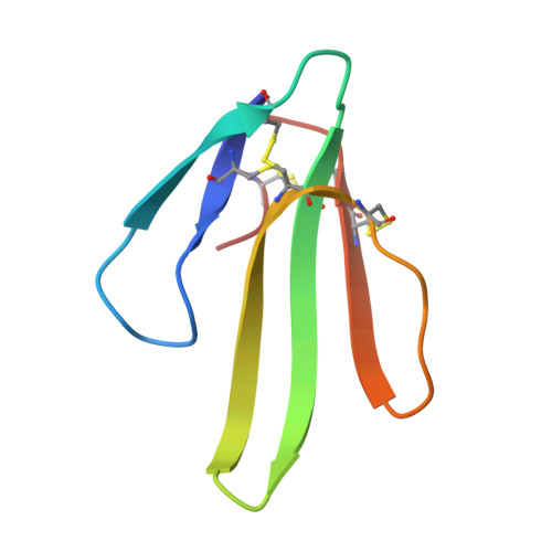
    The three-dimensional structure of erabutoxin a, a single-chain, 62-residue protein neurotoxin from snake venom, has been determined to 2.0-A resolution by x-ray crystal structure analysis. Molecular replacement methods were used, and the structure refined to a residual R = 0.17. The sites of 62 water molecules and 1 sulfate ion have been located and refined. The structure of erabutoxin a is very similar to that established earlier for erabutoxin b. These toxins from venom of the same snake differ in sequence only at residue 26, which is Asn in erabutoxin a and His in erabutoxin b. The substitution leads to only minor variations in intramolecular hydrogen bonding. Furthermore, the distribution of thermal parameters and the implied regional mobilities are similar in the two structures. In particular, the highly mobile character of the peripheral segment Pro44-Gly49 in both structures supports the specific role proposed for this segment in neurotoxin binding to the acetylcholine receptor. Forty-eight of the solvent sites determined are first surface positions; approximately one-half of these are equivalent to solvent sites in erabutoxin b.


  • Organizational Affiliation

    Department of Biochemistry and Molecular Biophysics, Columbia University, New York 10032.


Macromolecules
Find similar proteins by:  (by identity cutoff)  |  3D Structure
Entity ID: 1
MoleculeChains Sequence LengthOrganismDetailsImage
ERABUTOXIN A62Laticauda semifasciataMutation(s): 0 
UniProt
Find proteins for P60775 (Laticauda semifasciata)
Explore P60775 
Go to UniProtKB:  P60775
Entity Groups  
Sequence Clusters30% Identity50% Identity70% Identity90% Identity95% Identity100% Identity
UniProt GroupP60775
Sequence Annotations
Expand
  • Reference Sequence
Small Molecules
Ligands 1 Unique
IDChains Name / Formula / InChI Key2D Diagram3D Interactions
SO4
Query on SO4

Download Ideal Coordinates CCD File 
B [auth A]SULFATE ION
O4 S
QAOWNCQODCNURD-UHFFFAOYSA-L
Experimental Data & Validation

Experimental Data

  • Method: X-RAY DIFFRACTION
  • Resolution: 2.00 Å
  • R-Value Observed: 0.168 
  • Space Group: P 21 21 21
Unit Cell:
Length ( Å )Angle ( ˚ )
a = 50.14α = 90
b = 46.97β = 90
c = 20.87γ = 90
Software Package:
Software NamePurpose
PROLSQrefinement

Structure Validation

View Full Validation Report


Entry History 

Deposition Data

Revision History  (Full details and data files)

  • Version 1.0: 1990-04-15
    Type: Initial release
  • Version 1.1: 2008-03-03
    Changes: Version format compliance
  • Version 1.2: 2011-07-13
    Changes: Version format compliance
  • Version 1.3: 2017-11-29
    Changes: Derived calculations, Other