5EA7

Crystal Structure of Inhibitor BMS-433771 in Complex with Prefusion RSV F Glycoprotein


Experimental Data Snapshot

  • Method: X-RAY DIFFRACTION
  • Resolution: 2.85 Å
  • R-Value Free: 0.241 
  • R-Value Work: 0.196 
  • R-Value Observed: 0.198 

wwPDB Validation   3D Report Full Report


Ligand Structure Quality Assessment 


This is version 1.3 of the entry. See complete history


Literature

Molecular mechanism of respiratory syncytial virus fusion inhibitors.

Battles, M.B.Langedijk, J.P.Furmanova-Hollenstein, P.Chaiwatpongsakorn, S.Costello, H.M.Kwanten, L.Vranckx, L.Vink, P.Jaensch, S.Jonckers, T.H.Koul, A.Arnoult, E.Peeples, M.E.Roymans, D.McLellan, J.S.

(2016) Nat Chem Biol 12: 87-93

  • DOI: https://doi.org/10.1038/nchembio.1982
  • Primary Citation of Related Structures:  
    5EA3, 5EA4, 5EA5, 5EA6, 5EA7, 5EA8

  • PubMed Abstract: 

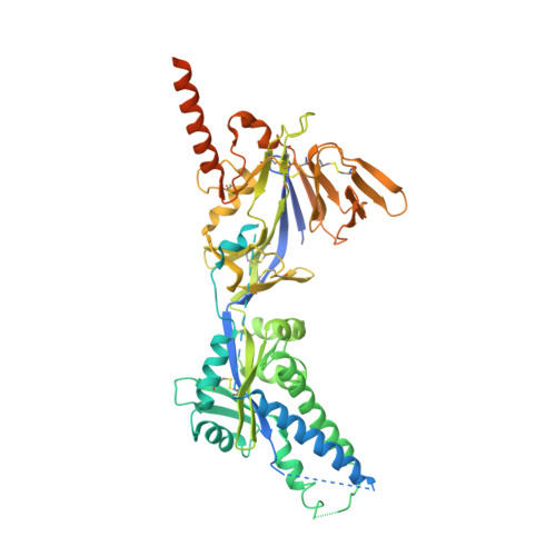
    Respiratory syncytial virus (RSV) is a leading cause of pneumonia and bronchiolitis in young children and the elderly. Therapeutic small molecules have been developed that bind the RSV F glycoprotein and inhibit membrane fusion, yet their binding sites and molecular mechanisms of action remain largely unknown. Here we show that these inhibitors bind to a three-fold-symmetric pocket within the central cavity of the metastable prefusion conformation of RSV F. Inhibitor binding stabilizes this conformation by tethering two regions that must undergo a structural rearrangement to facilitate membrane fusion. Inhibitor-escape mutations occur in residues that directly contact the inhibitors or are involved in the conformational rearrangements required to accommodate inhibitor binding. Resistant viruses do not propagate as well as wild-type RSV in vitro, indicating a fitness cost for inhibitor escape. Collectively, these findings provide new insight into class I viral fusion proteins and should facilitate development of optimal RSV fusion inhibitors.


  • Organizational Affiliation

    Department of Biochemistry, Geisel School of Medicine at Dartmouth, Hanover, New Hampshire, USA.


Macromolecules
Find similar proteins by:  (by identity cutoff)  |  3D Structure
Entity ID: 1
MoleculeChains Sequence LengthOrganismDetailsImage
Fusion glycoprotein F0A [auth F]568Human respiratory syncytial virus A2Mutation(s): 7 
UniProt
Find proteins for P03420 (Human respiratory syncytial virus A (strain A2))
Explore P03420 
Go to UniProtKB:  P03420
Entity Groups  
Sequence Clusters30% Identity50% Identity70% Identity90% Identity95% Identity100% Identity
UniProt GroupP03420
Sequence Annotations
Expand
  • Reference Sequence
Small Molecules
Binding Affinity Annotations 
IDSourceBinding Affinity
5NO BindingDB:  5EA7 EC50: min: 7.9, max: 28 (nM) from 3 assay(s)
Experimental Data & Validation

Experimental Data

Unit Cell:
Length ( Å )Angle ( ˚ )
a = 169.59α = 90
b = 169.59β = 90
c = 169.59γ = 90
Software Package:
Software NamePurpose
PHENIXrefinement
Aimlessdata scaling
PDB_EXTRACTdata extraction
iMOSFLMdata reduction
PHASERphasing

Structure Validation

View Full Validation Report


Ligand Structure Quality Assessment 


Entry History & Funding Information

Deposition Data


Funding OrganizationLocationGrant Number
Charles H. Hood FoundationUnited States--

Revision History  (Full details and data files)

  • Version 1.0: 2015-12-09
    Type: Initial release
  • Version 1.1: 2015-12-23
    Changes: Database references
  • Version 1.2: 2016-02-03
    Changes: Database references
  • Version 1.3: 2023-09-27
    Changes: Data collection, Database references, Derived calculations, Refinement description