5E8O

The structure of the TEIPP associated altered peptide ligand Trh4-p2ABU in complex with H-2D(b)


Experimental Data Snapshot

  • Method: X-RAY DIFFRACTION
  • Resolution: 1.98 Å
  • R-Value Free: 0.254 
  • R-Value Work: 0.218 
  • R-Value Observed: 0.220 

wwPDB Validation   3D Report Full Report


This is version 1.3 of the entry. See complete history


Literature

The MHC Class I Cancer-Associated Neoepitope Trh4 Linked with Impaired Peptide Processing Induces a Unique Noncanonical TCR Conformer.

Hafstrand, I.Doorduijn, E.M.Duru, A.D.Buratto, J.Oliveira, C.C.Sandalova, T.van Hall, T.Achour, A.

(2016) J Immunol 196: 2327-2334

  • DOI: https://doi.org/10.4049/jimmunol.1502249
  • Primary Citation of Related Structures:  
    5E8N, 5E8O, 5E8P

  • PubMed Abstract: 

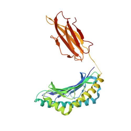
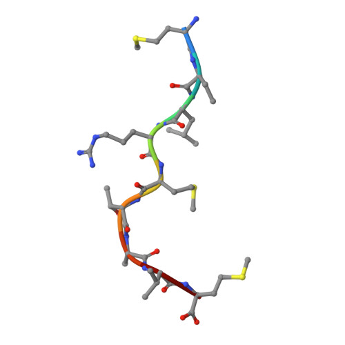
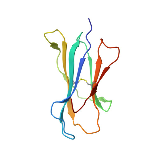
    MHC class I downregulation represents a significant challenge for successful T cell-based immunotherapy. T cell epitopes associated with impaired peptide processing (TEIPP) constitute a novel category of immunogenic Ags that are selectively presented on transporter associated with Ag processing-deficient cells. The TEIPP neoepitopes are CD8 T cell targets, derived from nonmutated self-proteins that might be exploited to prevent immune escape. In this study, the crystal structure of H-2D(b) in complex with the first identified TEIPP Ag (MCLRMTAVM) derived from the Trh4 protein has been determined to 2.25 Å resolution. In contrast to prototypic H-2D(b) peptides, Trh4 takes a noncanonical peptide-binding pattern with extensive sulfur-π interactions that contribute to the overall complex stability. Importantly, the noncanonical methionine at peptide position 5 acts as a main anchor, altering only the conformation of the H-2D(b) residues Y156 and H155 and thereby forming a unique MHC/peptide conformer that is essential for recognition by TEIPP-specific T cells. Substitution of peptide residues p2C and p5M to the conservative α-aminobutyric acid and norleucine, respectively, significantly reduced complex stability, without altering peptide conformation or T cell recognition. In contrast, substitution of p5M to a conventional asparagine abolished recognition by the H-2D(b)/Trh4-specific T cell clone LnB5. We anticipate that the H-2D(b)/Trh4 complex represents the first example, to our knowledge, of a broader repertoire of alternative MHC class I binders.


  • Organizational Affiliation

    Science for Life Laboratory, Department of Medicine, Solna, Karolinska Institutet, SE-10450 Stockholm, Sweden; and.


Macromolecules
Find similar proteins by:  (by identity cutoff)  |  3D Structure
Entity ID: 1
MoleculeChains Sequence LengthOrganismDetailsImage
H-2 class I histocompatibility antigen, D-B alpha chain
A, D
276Mus musculusMutation(s): 0 
Gene Names: H2-D1
UniProt
Find proteins for P01899 (Mus musculus)
Explore P01899 
Go to UniProtKB:  P01899
Entity Groups  
Sequence Clusters30% Identity50% Identity70% Identity90% Identity95% Identity100% Identity
UniProt GroupP01899
Sequence Annotations
Expand
  • Reference Sequence

Find similar proteins by:  Sequence   |   3D Structure  

Entity ID: 2
MoleculeChains Sequence LengthOrganismDetailsImage
Ceramide synthase 5B [auth C],
F
9Mus musculusMutation(s): 1 
EC: 2.3.1.24
UniProt & NIH Common Fund Data Resources
Find proteins for Q9D6K9 (Mus musculus)
Explore Q9D6K9 
Go to UniProtKB:  Q9D6K9
IMPC:  MGI:1919199
Entity Groups  
UniProt GroupQ9D6K9
Sequence Annotations
Expand
  • Reference Sequence
Find similar proteins by:  (by identity cutoff)  |  3D Structure
Entity ID: 3
MoleculeChains Sequence LengthOrganismDetailsImage
Beta-2-microglobulinC [auth B],
E
99Mus musculusMutation(s): 0 
Gene Names: B2m
UniProt & NIH Common Fund Data Resources
Find proteins for P01887 (Mus musculus)
Explore P01887 
Go to UniProtKB:  P01887
IMPC:  MGI:88127
Entity Groups  
Sequence Clusters30% Identity50% Identity70% Identity90% Identity95% Identity100% Identity
UniProt GroupP01887
Sequence Annotations
Expand
  • Reference Sequence
Small Molecules
Modified Residues  1 Unique
IDChains TypeFormula2D DiagramParent
ABA
Query on ABA
B [auth C],
F
L-PEPTIDE LINKINGC4 H9 N O2ALA
Experimental Data & Validation

Experimental Data

  • Method: X-RAY DIFFRACTION
  • Resolution: 1.98 Å
  • R-Value Free: 0.254 
  • R-Value Work: 0.218 
  • R-Value Observed: 0.220 
  • Space Group: I 1 2 1
Unit Cell:
Length ( Å )Angle ( ˚ )
a = 90.859α = 90
b = 123.33β = 104.67
c = 97.552γ = 90
Software Package:
Software NamePurpose
PHENIXrefinement
MOSFLMdata reduction
Aimlessdata scaling
PHASERphasing

Structure Validation

View Full Validation Report


Entry History 

Deposition Data

Revision History  (Full details and data files)

  • Version 1.0: 2016-02-03
    Type: Initial release
  • Version 1.1: 2016-02-10
    Changes: Database references
  • Version 1.2: 2016-03-02
    Changes: Database references
  • Version 1.3: 2024-01-10
    Changes: Data collection, Database references, Refinement description