5E5J

Joint X-ray/neutron structure of HIV-1 protease triple mutant (V32I,I47V,V82I) with darunavir at pH 6.0


Experimental Data Snapshot

  • Method: X-RAY DIFFRACTION
  • Resolution: 1.85 Å
  • R-Value Free: 0.201 
  • R-Value Work: 0.194 

  • Method: NEUTRON DIFFRACTION
  • Resolution: 2.00 Å
  • R-Value Free: 0.245 
  • R-Value Work: 0.217 

wwPDB Validation   3D Report Full Report


This is version 1.1 of the entry. See complete history


Literature

Long-Range Electrostatics-Induced Two-Proton Transfer Captured by Neutron Crystallography in an Enzyme Catalytic Site.

Gerlits, O.Wymore, T.Das, A.Shen, C.H.Parks, J.M.Smith, J.C.Weiss, K.L.Keen, D.A.Blakeley, M.P.Louis, J.M.Langan, P.Weber, I.T.Kovalevsky, A.

(2016) Angew Chem Int Ed Engl 55: 4924-4927

  • DOI: https://doi.org/10.1002/anie.201509989
  • Primary Citation of Related Structures:  
    5E5J, 5E5K

  • PubMed Abstract: 

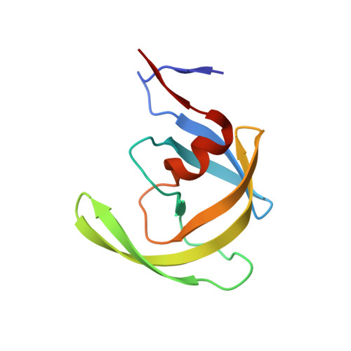
    Neutron crystallography was used to directly locate two protons before and after a pH-induced two-proton transfer between catalytic aspartic acid residues and the hydroxy group of the bound clinical drug darunavir, located in the catalytic site of enzyme HIV-1 protease. The two-proton transfer is triggered by electrostatic effects arising from protonation state changes of surface residues far from the active site. The mechanism and pH effect are supported by quantum mechanics/molecular mechanics (QM/MM) calculations. The low-pH proton configuration in the catalytic site is deemed critical for the catalytic action of this enzyme and may apply more generally to other aspartic proteases. Neutrons therefore represent a superb probe to obtain structural details for proton transfer reactions in biological systems at a truly atomic level.


  • Organizational Affiliation

    Biology and Soft Matter Division, Oak Ridge National Laboratory, Oak Ridge, TN, 37831, USA.


Macromolecules
Find similar proteins by:  (by identity cutoff)  |  3D Structure
Entity ID: 1
MoleculeChains Sequence LengthOrganismDetailsImage
Protease
A, B
99Human immunodeficiency virus 1Mutation(s): 8 
Gene Names: pol
UniProt
Find proteins for Q7SSI0 (Human immunodeficiency virus 1)
Explore Q7SSI0 
Go to UniProtKB:  Q7SSI0
Entity Groups  
Sequence Clusters30% Identity50% Identity70% Identity90% Identity95% Identity100% Identity
UniProt GroupQ7SSI0
Sequence Annotations
Expand
  • Reference Sequence
Small Molecules
Ligands 1 Unique
IDChains Name / Formula / InChI Key2D Diagram3D Interactions
017
Query on 017

Download Ideal Coordinates CCD File 
C [auth A](3R,3AS,6AR)-HEXAHYDROFURO[2,3-B]FURAN-3-YL(1S,2R)-3-[[(4-AMINOPHENYL)SULFONYL](ISOBUTYL)AMINO]-1-BENZYL-2-HYDROXYPROPYLCARBAMATE
C27 H37 N3 O7 S
CJBJHOAVZSMMDJ-HEXNFIEUSA-N
Binding Affinity Annotations 
IDSourceBinding Affinity
017 BindingDB:  5E5J Ki: min: 2.00e-4, max: 2 (nM) from 16 assay(s)
Kd: 0.02 (nM) from 1 assay(s)
IC50: min: 0.06, max: 370 (nM) from 10 assay(s)
EC50: min: 0.25, max: 112 (nM) from 4 assay(s)
Experimental Data & Validation

Experimental Data

  • Method: X-RAY DIFFRACTION
  • Resolution: 1.85 Å
  • R-Value Free: 0.201 
  • R-Value Work: 0.194 
  • Space Group: P 21 21 2
  • Method: NEUTRON DIFFRACTION
  • Resolution: 2.00 Å
  • R-Value Free: 0.245 
  • R-Value Work: 0.217 
  • Space Group: P 21 21 2
Unit Cell:
Length ( Å )Angle ( ˚ )
a = 59.724α = 90
b = 87.257β = 90
c = 46.547γ = 90
Software Package:
Software NamePurpose
HKL-3000data reduction
HKL-3000data scaling
PHASERphasing
nCNSrefinement
LAUEGENdata reduction
LSCALEdata scaling

Structure Validation

View Full Validation Report


Entry History 

Deposition Data

Revision History  (Full details and data files)

  • Version 1.0: 2016-05-04
    Type: Initial release
  • Version 1.1: 2024-03-06
    Changes: Data collection, Database references, Derived calculations, Structure summary