5DYO

Fab43.1 complex with flourescein


Experimental Data Snapshot

  • Method: X-RAY DIFFRACTION
  • Resolution: 2.36 Å
  • R-Value Free: 0.239 
  • R-Value Work: 0.193 
  • R-Value Observed: 0.196 

wwPDB Validation   3D Report Full Report


Ligand Structure Quality Assessment 


This is version 2.2 of the entry. See complete history


Literature

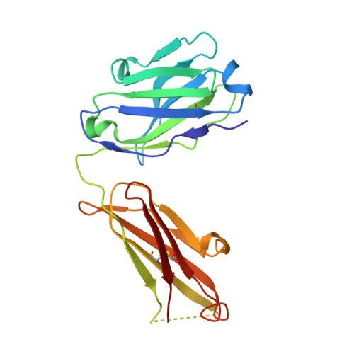
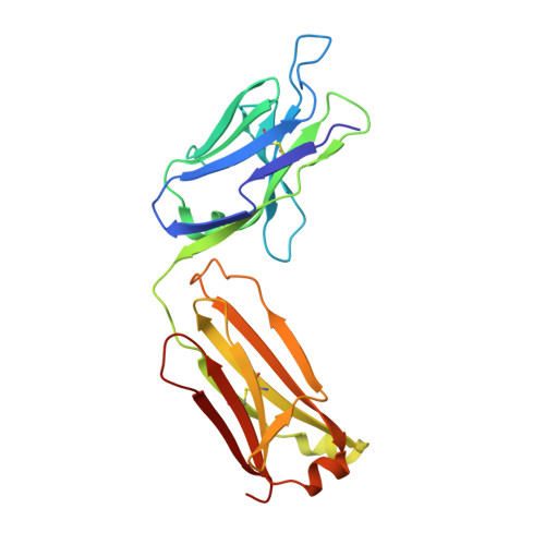
Three-dimensional structure, binding, and spectroscopic characteristics of the monoclonal antibody 43.1 directed to the carboxyphenyl moiety of fluorescein.

Gayda, S.Longenecker, K.L.Judge, R.A.Swift, K.M.Manoj, S.Linthicum, D.S.Tetin, S.Y.

(2016) Biopolymers 105: 234-243

  • DOI: https://doi.org/10.1002/bip.22801
  • Primary Citation of Related Structures:  
    5DYO

  • PubMed Abstract: 

    Unlike other known anti-fluorescein antibodies, the monoclonal antibody 43.1 is directed toward the fluorescein's carboxyl phenyl moiety. It demonstrates a very high affinity (KD ∼ 70 pM) and a fast association rate (kon ∼ 2 × 10(7) M(-1 ) s(-1) ). The three-dimensional structure of the Fab 43.1-fluorescein complex was resolved at 2.4 Å resolution. The antibody binding site is exclusively assembled by the CDR loops. It is comprised of a 14 Å groove-shaped entrance leading to a 9 Å by 7 Å binding pocket. The highly polar binding pocket complementary encloses the fluorescein's carboxyphenyl moiety and tightly fixes it by multiple hydrogen bonds. The fluorescein's xanthene ring is embedded in the more hydrophobic groove and stacked between the side chains of Tyr37L and of Arg99H providing conditions for an excited state electron transfer process. In comparison to fluorescein, the absorption spectrum of the complex in the visible region is shifted to the "red" by 23 nm. The complex demonstrates a very weak fluorescence (Φc  = 0.0018) with two short lifetime components: 0.03 ns (47%) and 0.8 ns (24%), which reflects a 99.8% fluorescein emission quenching effect upon complex formation. The antibody 43.1 binds fluorescein with remarkable affinity, fast association rate, and strongly quenches its emission. Therefore, it may present a practical interest in applications such as molecular sensors and switches.


  • Organizational Affiliation

    Diagnostics Research, Abbott Diagnostics Division, Abbott Park, IL, 60064.


Macromolecules
Find similar proteins by:  (by identity cutoff)  |  3D Structure
Entity ID: 1
MoleculeChains Sequence LengthOrganismDetailsImage
Fab 43.1 Heavy ChainA,
C [auth H]
212Mus musculusMutation(s): 0 
Entity Groups  
Sequence Clusters30% Identity50% Identity70% Identity90% Identity95% Identity100% Identity
Sequence Annotations
Expand
  • Reference Sequence
Find similar proteins by:  (by identity cutoff)  |  3D Structure
Entity ID: 2
MoleculeChains Sequence LengthOrganismDetailsImage
Fab 43.1 Light ChainB,
D [auth L]
218Mus musculusMutation(s): 0 
Entity Groups  
Sequence Clusters30% Identity50% Identity70% Identity90% Identity95% Identity100% Identity
Sequence Annotations
Expand
  • Reference Sequence
Experimental Data & Validation

Experimental Data

  • Method: X-RAY DIFFRACTION
  • Resolution: 2.36 Å
  • R-Value Free: 0.239 
  • R-Value Work: 0.193 
  • R-Value Observed: 0.196 
  • Space Group: P 1 21 1
Unit Cell:
Length ( Å )Angle ( ˚ )
a = 54.232α = 90
b = 105.401β = 102.73
c = 109.059γ = 90
Software Package:
Software NamePurpose
BUSTER-TNTrefinement
PDB_EXTRACTdata extraction
XDSdata reduction
Aimlessdata scaling
MOLREPphasing

Structure Validation

View Full Validation Report


Ligand Structure Quality Assessment 


Entry History 

Deposition Data

Revision History  (Full details and data files)

  • Version 1.0: 2016-01-27
    Type: Initial release
  • Version 1.1: 2016-02-10
    Changes: Database references
  • Version 2.0: 2017-09-20
    Changes: Atomic model, Database references, Derived calculations, Non-polymer description, Structure summary
  • Version 2.1: 2018-03-21
    Changes: Data collection
  • Version 2.2: 2023-09-27
    Changes: Data collection, Database references, Refinement description