5DRN

Context-independent anti-hypusine antibody FabHpu24 in complex with hypusine


Experimental Data Snapshot

  • Method: X-RAY DIFFRACTION
  • Resolution: 1.99 Å
  • R-Value Free: 0.222 
  • R-Value Work: 0.183 
  • R-Value Observed: 0.185 

wwPDB Validation   3D Report Full Report


Ligand Structure Quality Assessment 


This is version 2.1 of the entry. See complete history


Literature

Structural Analysis and Optimization of Context-Independent Anti-Hypusine Antibodies.

Zhai, Q.He, M.Song, A.Deshayes, K.Dixit, V.M.Carter, P.J.

(2016) J Mol Biol 428: 603-617

  • DOI: https://doi.org/10.1016/j.jmb.2016.01.006
  • Primary Citation of Related Structures:  
    5DRN, 5DS8, 5DSC, 5DTF, 5DUB

  • PubMed Abstract: 

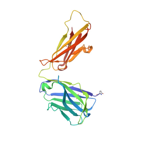
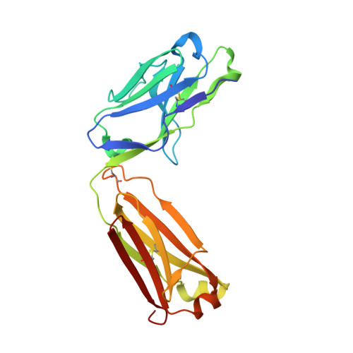
    Context-independent anti-hypusine antibodies that bind to the post-translational modification (PTM), hypusine, with minimal dependence on flanking amino acid sequences, were identified. The antibodies bind to both hypusine and deoxyhypusine or selectively to hypusine but not to deoxyhypusine. Phage display was used to further enhance the affinity of the antibodies. Affinity maturation of these anti-hypusine antibodies improved their performance in affinity capture of the only currently known hypusinated protein, eukaryotic translation initiation factor 5A. These anti-hypusine antibodies may have utility in the identification of novel hypusinated proteins. Crystal structures of the corresponding Fab fragments were determined in complex with hypusine- or deoxyhypusine-containing peptides. The hypusine or deoxyhypusine moiety was found to reside in a deep pocket formed between VH and VL domains of the Fab fragments. Interaction between the antibodies and hypusine includes an extensive hydrogen bond network. These are, to our knowledge, the first reported structures of context-independent anti-PTM antibodies in complex with the corresponding PTM.


  • Organizational Affiliation

    Department of Antibody Engineering, Genentech Inc., 1 DNA Way, South San Francisco, CA 94080, USA.


Macromolecules
Find similar proteins by:  (by identity cutoff)  |  3D Structure
Entity ID: 1
MoleculeChains Sequence LengthOrganismDetailsImage
Fab Hpu24 Heavy chainA,
C [auth H]
216Oryctolagus cuniculusMutation(s): 0 
Entity Groups  
Sequence Clusters30% Identity50% Identity70% Identity90% Identity95% Identity100% Identity
Sequence Annotations
Expand
  • Reference Sequence
Find similar proteins by:  (by identity cutoff)  |  3D Structure
Entity ID: 2
MoleculeChains Sequence LengthOrganismDetailsImage
Fab Hpu24 Light chainB,
D [auth L]
215Oryctolagus cuniculusMutation(s): 0 
Entity Groups  
Sequence Clusters30% Identity50% Identity70% Identity90% Identity95% Identity100% Identity
Sequence Annotations
Expand
  • Reference Sequence
Experimental Data & Validation

Experimental Data

  • Method: X-RAY DIFFRACTION
  • Resolution: 1.99 Å
  • R-Value Free: 0.222 
  • R-Value Work: 0.183 
  • R-Value Observed: 0.185 
  • Space Group: P 1 21 1
Unit Cell:
Length ( Å )Angle ( ˚ )
a = 42.453α = 90
b = 167.35β = 98.93
c = 68.326γ = 90
Software Package:
Software NamePurpose
SCALEPACKdata scaling
PHENIXrefinement
PDB_EXTRACTdata extraction
PHASERphasing
HKL-2000data reduction

Structure Validation

View Full Validation Report


Ligand Structure Quality Assessment 


Entry History 

Deposition Data

Revision History  (Full details and data files)

  • Version 1.0: 2016-01-20
    Type: Initial release
  • Version 1.1: 2016-02-03
    Changes: Database references
  • Version 1.2: 2016-03-02
    Changes: Database references
  • Version 1.3: 2016-05-04
    Changes: Non-polymer description
  • Version 2.0: 2023-09-27
    Changes: Data collection, Database references, Derived calculations, Polymer sequence, Refinement description
  • Version 2.1: 2023-11-15
    Changes: Data collection