5DFW

CRYSTAL STRUCTURE OF HUMAN CD81 LARGE EXTRACELLULAR LOOP IN COMPLEX WITH SINGLE CHAIN FV FRAGMENT K13


Experimental Data Snapshot

  • Method: X-RAY DIFFRACTION
  • Resolution: 2.33 Å
  • R-Value Free: 0.264 
  • R-Value Work: 0.190 
  • R-Value Observed: 0.194 

wwPDB Validation   3D Report Full Report


This is version 1.1 of the entry. See complete history


Literature

VH-VL orientation prediction for antibody humanization candidate selection: A case study.

Bujotzek, A.Lipsmeier, F.Harris, S.F.Benz, J.Kuglstatter, A.Georges, G.

(2016) MAbs 8: 288-305

  • DOI: https://doi.org/10.1080/19420862.2015.1117720
  • Primary Citation of Related Structures:  
    5DFV, 5DFW, 5DMG

  • PubMed Abstract: 

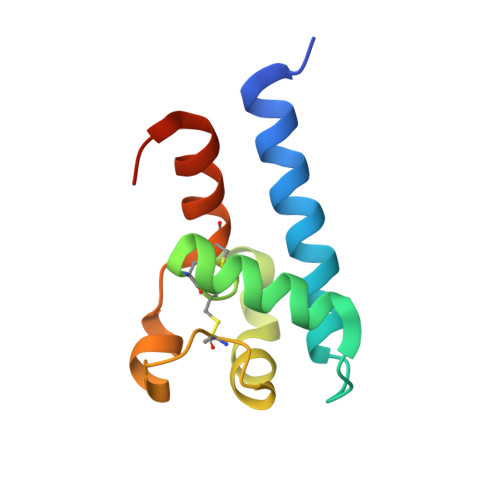
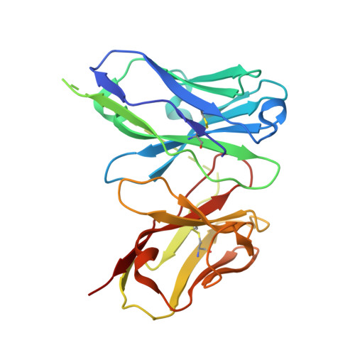
    Antibody humanization describes the procedure of grafting a non-human antibody's complementarity-determining regions, i.e., the variable loop regions that mediate specific interactions with the antigen, onto a β-sheet framework that is representative of the human variable region germline repertoire, thus reducing the number of potentially antigenic epitopes that might trigger an anti-antibody response. The selection criterion for the so-called acceptor frameworks (one for the heavy and one for the light chain variable region) is traditionally based on sequence similarity. Here, we propose a novel approach that selects acceptor frameworks such that the relative orientation of the 2 variable domains in 3D space, and thereby the geometry of the antigen-binding site, is conserved throughout the process of humanization. The methodology relies on a machine learning-based predictor of antibody variable domain orientation that has recently been shown to improve the quality of antibody homology models. Using data from 3 humanization campaigns, we demonstrate that preselecting humanization variants based on the predicted difference in variable domain orientation with regard to the original antibody leads to subsets of variants with a significant improvement in binding affinity.


  • Organizational Affiliation

    a Roche Pharmaceutical Research and Early Development, Large Molecule Research, Roche Innovation Center Penzberg , Nonnenwald 2, Penzberg , Germany.


Macromolecules
Find similar proteins by:  (by identity cutoff)  |  3D Structure
Entity ID: 1
MoleculeChains Sequence LengthOrganismDetailsImage
CD81 antigen98Homo sapiensMutation(s): 0 
Gene Names: CD81TAPA1TSPAN28
UniProt & NIH Common Fund Data Resources
Find proteins for P60033 (Homo sapiens)
Explore P60033 
Go to UniProtKB:  P60033
PHAROS:  P60033
GTEx:  ENSG00000110651 
Entity Groups  
Sequence Clusters30% Identity50% Identity70% Identity90% Identity95% Identity100% Identity
UniProt GroupP60033
Sequence Annotations
Expand
  • Reference Sequence
Find similar proteins by:  (by identity cutoff)  |  3D Structure
Entity ID: 2
MoleculeChains Sequence LengthOrganismDetailsImage
SINGLE CHAIN FV FRAGMENTB [auth H]243Mus musculusMutation(s): 0 
Entity Groups  
Sequence Clusters30% Identity50% Identity70% Identity90% Identity95% Identity100% Identity
Sequence Annotations
Expand
  • Reference Sequence
Experimental Data & Validation

Experimental Data

  • Method: X-RAY DIFFRACTION
  • Resolution: 2.33 Å
  • R-Value Free: 0.264 
  • R-Value Work: 0.190 
  • R-Value Observed: 0.194 
  • Space Group: P 1 2 1
Unit Cell:
Length ( Å )Angle ( ˚ )
a = 49.652α = 90
b = 49.671β = 105.3
c = 80.452γ = 90
Software Package:
Software NamePurpose
BUSTERrefinement
SCALEPACKdata scaling
DENZOdata reduction
PHASERphasing

Structure Validation

View Full Validation Report


Entry History 

Deposition Data

Revision History  (Full details and data files)

  • Version 1.0: 2015-12-16
    Type: Initial release
  • Version 1.1: 2016-03-02
    Changes: Database references