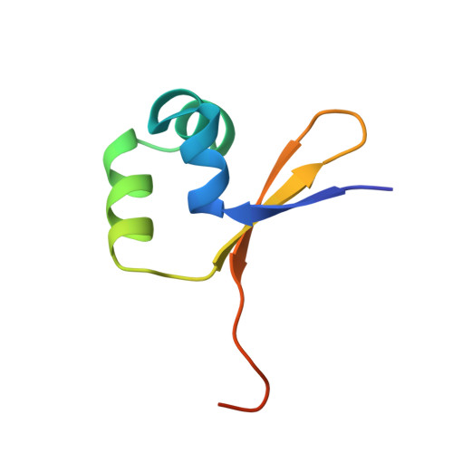
Refined structure of Cro repressor protein from bacteriophage lambda suggests both flexibility and plasticity.
Ohlendorf, D.H., Tronrud, D.E., Matthews, B.W.(1998) J Mol Biol 280: 129-136
- PubMed: 9653036
- DOI: https://doi.org/10.1006/jmbi.1998.1849
- Primary Citation of Related Structures:
5CRO - PubMed Abstract:
The structure of the Cro repressor protein from phage lambda has been refined to a crystallographic R-value of 19.3% at 2.3 A resolution. The re fined model supports the structure as originally described in 1981 and provides a basis for comparison with the Cro-operator complex described in the accompanying paper. Changes in structure seen in different crystal forms and modifications of Cro suggest that the individual subunits are somewhat plastic in nature. In addition, the dimer of Cro suggests a high degree of flexibility, which may be important in forming the Cro-DNA complex. The structure of the Cro subunit as determined by NMR agrees reasonably well with that in the crystals (root-mean-square discrepancy of about 2 A for all atoms). There are, however, only a limited number of intersubunit distance constraints and, presumably for this reason, the different NMR models for the dimer vary substantially among themselves (discrepancies of 1.3 to 5.5 A). Because of this variation it is not possible to say whether the range of discrepancies between the X-ray and NMR Cro dimers (2.9 to 7.5 A) represent a significant difference between the X-ray and solution structures. It has previously been proposed that substitutions of Tyr26 in Cro increase thermal stability by the "reverse hydrophobic effect", i.e. by exposing 40% more hydrophobic surface to solvent in the folded form than in the unfolded state. The refined structure, however, suggests that Tyr26 is equally solvent exposed in the folded and unfolded states. The most stabilizing substitution is Tyr26-->Asp and in this case it appears that interaction with an alpha-helix dipole is at least partly responsible for the enhanced stability.
Organizational Affiliation:
Institute of Molecular Biology Howard Hughes Medical Institute and Department of Physics, University of Oregon, Eugene, OR, 97403-1229, USA.