5C69

Crystal Structure of Prefusion-stabilized RSV F variant PR-DM


Experimental Data Snapshot

  • Method: X-RAY DIFFRACTION
  • Resolution: 2.30 Å
  • R-Value Free: 0.217 
  • R-Value Work: 0.180 
  • R-Value Observed: 0.182 

wwPDB Validation   3D Report Full Report


This is version 1.2 of the entry. See complete history


Literature

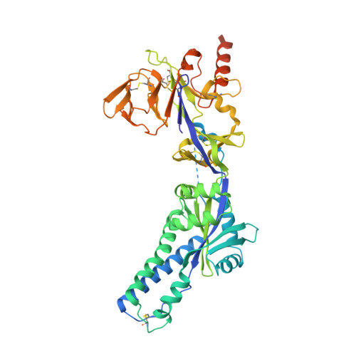
A highly stable prefusion RSV F vaccine derived from structural analysis of the fusion mechanism.

Krarup, A.Truan, D.Furmanova-Hollenstein, P.Bogaert, L.Bouchier, P.Bisschop, I.J.Widjojoatmodjo, M.N.Zahn, R.Schuitemaker, H.McLellan, J.S.Langedijk, J.P.

(2015) Nat Commun 6: 8143-8143

  • DOI: https://doi.org/10.1038/ncomms9143
  • Primary Citation of Related Structures:  
    5C69, 5C6B

  • PubMed Abstract: 

    Respiratory syncytial virus (RSV) causes acute lower respiratory tract infections and is the leading cause of infant hospitalizations. Recently, a promising vaccine antigen based on the RSV fusion protein (RSV F) stabilized in the native prefusion conformation has been described. Here we report alternative strategies to arrest RSV F in the prefusion conformation based on the prevention of hinge movements in the first refolding region and the elimination of proteolytic exposure of the fusion peptide. A limited number of unique mutations are identified that stabilize the prefusion conformation of RSV F and dramatically increase expression levels. This highly stable prefusion RSV F elicits neutralizing antibodies in cotton rats and induces complete protection against viral challenge. Moreover, the structural and biochemical analysis of the prefusion variants suggests a function for p27, the excised segment that precedes the fusion peptide in the polypeptide chain.


  • Organizational Affiliation

    Janssen Infectious Diseases and Vaccines, Archimedesweg 4-6, Leiden 2333 CN, The Netherlands.


Macromolecules
Find similar proteins by:  (by identity cutoff)  |  3D Structure
Entity ID: 1
MoleculeChains Sequence LengthOrganismDetailsImage
Fusion glycoprotein F0,Fibritin492Human respiratory syncytial virus A2Enterobacteria phage Ox2
This entity is chimeric
Mutation(s): 4 
UniProt
Find proteins for Q38650 (Enterobacteria phage Ox2)
Explore Q38650 
Go to UniProtKB:  Q38650
Find proteins for P03420 (Human respiratory syncytial virus A (strain A2))
Explore P03420 
Go to UniProtKB:  P03420
Entity Groups  
Sequence Clusters30% Identity50% Identity70% Identity90% Identity95% Identity100% Identity
UniProt GroupsP03420Q38650
Sequence Annotations
Expand
  • Reference Sequence
Small Molecules
Ligands 3 Unique
IDChains Name / Formula / InChI Key2D Diagram3D Interactions
NHE
Query on NHE

Download Ideal Coordinates CCD File 
I [auth A],
J [auth A]
2-[N-CYCLOHEXYLAMINO]ETHANE SULFONIC ACID
C8 H17 N O3 S
MKWKNSIESPFAQN-UHFFFAOYSA-N
SO4
Query on SO4

Download Ideal Coordinates CCD File 
B [auth A]
C [auth A]
D [auth A]
E [auth A]
F [auth A]
B [auth A],
C [auth A],
D [auth A],
E [auth A],
F [auth A],
G [auth A],
H [auth A]
SULFATE ION
O4 S
QAOWNCQODCNURD-UHFFFAOYSA-L
CL
Query on CL

Download Ideal Coordinates CCD File 
K [auth A],
L [auth A],
M [auth A],
N [auth A],
O [auth A]
CHLORIDE ION
Cl
VEXZGXHMUGYJMC-UHFFFAOYSA-M
Experimental Data & Validation

Experimental Data

Unit Cell:
Length ( Å )Angle ( ˚ )
a = 167.82α = 90
b = 167.82β = 90
c = 167.82γ = 90
Software Package:
Software NamePurpose
Aimlessdata scaling
PHASERphasing
PHENIXrefinement
PDB_EXTRACTdata extraction
Cootmodel building
iMOSFLMdata reduction

Structure Validation

View Full Validation Report


Entry History 

Deposition Data

Revision History  (Full details and data files)

  • Version 1.0: 2015-09-23
    Type: Initial release
  • Version 1.1: 2021-03-24
    Changes: Derived calculations, Source and taxonomy
  • Version 1.2: 2023-09-27
    Changes: Data collection, Database references, Refinement description