5BTW

Structure of the N-terminal domain of lpg1496 from Legionella pneumophila


Experimental Data Snapshot

  • Method: X-RAY DIFFRACTION
  • Resolution: 1.20 Å
  • R-Value Free: 0.180 
  • R-Value Work: 0.158 
  • R-Value Observed: 0.159 

wwPDB Validation   3D Report Full Report


This is version 1.5 of the entry. See complete history


Literature

Structure of the Legionella Effector, lpg1496, Suggests a Role in Nucleotide Metabolism.

Wong, K.Kozlov, G.Zhang, Y.Gehring, K.

(2015) J Biol Chem 290: 24727-24737

  • DOI: https://doi.org/10.1074/jbc.M115.671263
  • Primary Citation of Related Structures:  
    5BTW, 5BTX, 5BTY, 5BTZ, 5BU0, 5BU1

  • PubMed Abstract: 

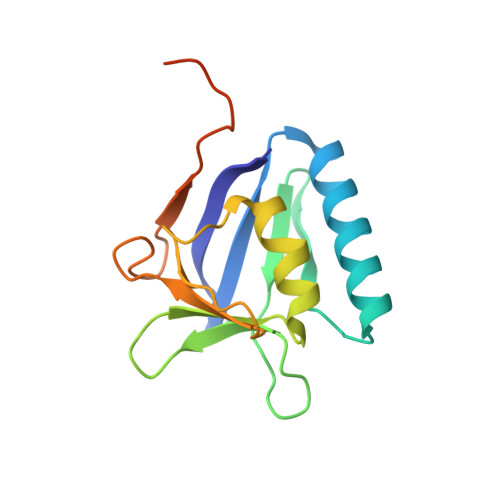
    Pathogenic Gram-negative bacteria use specialized secretion systems that translocate bacterial proteins, termed effectors, directly into host cells where they interact with host proteins and biochemical processes for the benefit of the pathogen. lpg1496 is a previously uncharacterized effector of Legionella pneumophila, the causative agent of Legionnaires disease. Here, we crystallized three nucleotide binding domains from lpg1496. The C-terminal domain, which is conserved among the SidE family of effectors, is formed of two largely α-helical lobes with a nucleotide binding cleft. A structural homology search has shown similarity to phosphodiesterases involved in cleavage of cyclic nucleotides. We have also crystallized a novel domain that occurs twice in the N-terminal half of the protein that we term the KLAMP domain due to the presence of homologous domains in bacterial histidine kinase-like ATP binding region-containing proteins and S-adenosylmethionine-dependent methyltransferase proteins. Both KLAMP structures are very similar but selectively bind 3',5'-cAMP and ADP. A co-crystal of the KLAMP1 domain with 3',5'-cAMP reveals the contribution of Tyr-61 and Tyr-69 that produces π-stacking interactions with the adenine ring of the nucleotide. Our study provides the first structural insights into two novel nucleotide binding domains associated with bacterial virulence.


  • Organizational Affiliation

    From the Department of Biochemistry and Groupe de Recherche Axé sur la Structure des Protéines, McGill University, Montreal, Quebec H3G 0B1, Canada.


Macromolecules
Find similar proteins by:  (by identity cutoff)  |  3D Structure
Entity ID: 1
MoleculeChains Sequence LengthOrganismDetailsImage
Uncharacterized protein
A, B
146Legionella pneumophila subsp. pneumophila str. Philadelphia 1Mutation(s): 0 
Gene Names: lpg1496
UniProt
Find proteins for Q5ZVE4 (Legionella pneumophila subsp. pneumophila (strain Philadelphia 1 / ATCC 33152 / DSM 7513))
Explore Q5ZVE4 
Go to UniProtKB:  Q5ZVE4
Entity Groups  
Sequence Clusters30% Identity50% Identity70% Identity90% Identity95% Identity100% Identity
UniProt GroupQ5ZVE4
Sequence Annotations
Expand
  • Reference Sequence
Experimental Data & Validation

Experimental Data

  • Method: X-RAY DIFFRACTION
  • Resolution: 1.20 Å
  • R-Value Free: 0.180 
  • R-Value Work: 0.158 
  • R-Value Observed: 0.159 
  • Space Group: P 1 21 1
Unit Cell:
Length ( Å )Angle ( ˚ )
a = 32.41α = 90
b = 56.92β = 91.18
c = 60.184γ = 90
Software Package:
Software NamePurpose
REFMACrefinement
HKL-2000data reduction
HKL-2000data scaling
ARPmodel building
Cootmodel building

Structure Validation

View Full Validation Report


Entry History & Funding Information

Deposition Data


Funding OrganizationLocationGrant Number
Canadian Institutes of Health Research (CIHR)CanadaGSP-48370

Revision History  (Full details and data files)

  • Version 1.0: 2015-08-26
    Type: Initial release
  • Version 1.1: 2015-09-02
    Changes: Database references
  • Version 1.2: 2015-10-21
    Changes: Database references
  • Version 1.3: 2017-09-06
    Changes: Author supporting evidence, Database references, Derived calculations
  • Version 1.4: 2020-01-08
    Changes: Author supporting evidence
  • Version 1.5: 2023-09-27
    Changes: Data collection, Database references, Experimental preparation, Refinement description