5BNY

Crystal structure of hemagglutinin of A/Chicken/Guangdong/S1311/2010 (H6N6)


Experimental Data Snapshot

  • Method: X-RAY DIFFRACTION
  • Resolution: 2.66 Å
  • R-Value Free: 0.245 
  • R-Value Work: 0.202 
  • R-Value Observed: 0.204 

wwPDB Validation   3D Report Full Report


Ligand Structure Quality Assessment 


This is version 2.1 of the entry. See complete history


Literature

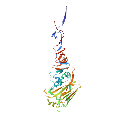
Structural and Functional Studies of Influenza Virus A/H6 Hemagglutinin.

Ni, F.Kondrashkina, E.Wang, Q.

(2015) PLoS One 7: e0134576-e0134576

  • DOI: https://doi.org/10.1371/journal.pone.0134576
  • Primary Citation of Related Structures:  
    5BNY, 5BQY, 5BQZ, 5BR0, 5BR3, 5BR6

  • PubMed Abstract: 

    In June 2013, the first human infection by avian influenza A(H6N1) virus was reported in Taiwan. This incident raised the concern for possible human epidemics and pandemics from H6 viruses. In this study, we performed structural and functional investigation on the hemagglutinin (HA) proteins of the human-infecting A/Taiwan/2/2013(H6N1) (TW H6) virus and an avian A/chicken/Guangdong/S1311/2010(H6N6) (GD H6) virus that transmitted efficiently in guinea pigs. Our results revealed that in the presence of HA1 Q226, the triad of HA1 S137, E190 and G228 in GD H6 HA allows the binding to both avian- and human-like receptors with a slight preference for avian receptors. Its conservation among the majority of H6 HAs provides an explanation for the broader host range of this subtype. Furthermore, the triad of N137, V190 and S228 in TW H6 HA may alleviate the requirement for a hydrophobic residue at HA1 226 of H2 and H3 HAs when binding to human-like receptors. Consequently, TW H6 HA has a slight preference for human receptors, thus may represent an intermediate towards a complete human adaptation. Importantly, the triad observed in TW H6 HA is detected in 74% H6 viruses isolated from Taiwan in the past 14 years, suggesting an elevated threat of H6 viruses from this region to human health. The novel roles of the triad at HA1 137, 190 and 228 of H6 HA in binding to receptors revealed here may also be used by other HA subtypes to achieve human adaptation, which needs to be further tested in laboratory and closely monitored in field surveillance.


  • Organizational Affiliation

    Verna and Marrs McLean Department of Biochemistry and Molecular Biology, Baylor College of Medicine, Houston, Texas, United States of America.


Macromolecules
Find similar proteins by:  (by identity cutoff)  |  3D Structure
Entity ID: 1
MoleculeChains Sequence LengthOrganismDetailsImage
Hemagglutinin
A, C, E
324Influenza A virus (A/chicken/Guangdong/S1311/2010(H6N6))Mutation(s): 0 
Gene Names: HA
UniProt
Find proteins for A0A067YZV9 (Influenza A virus)
Explore A0A067YZV9 
Go to UniProtKB:  A0A067YZV9
Entity Groups  
Sequence Clusters30% Identity50% Identity70% Identity90% Identity95% Identity100% Identity
UniProt GroupA0A067YZV9
Sequence Annotations
Expand
  • Reference Sequence
Find similar proteins by:  (by identity cutoff)  |  3D Structure
Entity ID: 2
MoleculeChains Sequence LengthOrganismDetailsImage
Hemagglutinin
B, D, F
191Influenza A virus (A/chicken/Guangdong/S1311/2010(H6N6))Mutation(s): 0 
Gene Names: HA
UniProt
Find proteins for A0A067YZV9 (Influenza A virus)
Explore A0A067YZV9 
Go to UniProtKB:  A0A067YZV9
Entity Groups  
Sequence Clusters30% Identity50% Identity70% Identity90% Identity95% Identity100% Identity
UniProt GroupA0A067YZV9
Sequence Annotations
Expand
  • Reference Sequence
Oligosaccharides

Help

Entity ID: 3
MoleculeChains Length2D Diagram Glycosylation3D Interactions
2-acetamido-2-deoxy-beta-D-glucopyranose-(1-4)-2-acetamido-2-deoxy-beta-D-glucopyranose
G, H, I, J
2N-Glycosylation
Glycosylation Resources
GlyTouCan:  G42666HT
GlyCosmos:  G42666HT
GlyGen:  G42666HT
Entity ID: 4
MoleculeChains Length2D Diagram Glycosylation3D Interactions
2-acetamido-2-deoxy-beta-D-glucopyranose-(1-4)-2-acetamido-2-deoxy-beta-D-glucopyranose-(1-4)-2-acetamido-2-deoxy-beta-D-glucopyranose
K
3N-Glycosylation
Glycosylation Resources
GlyTouCan:  G47362BJ
GlyCosmos:  G47362BJ
GlyGen:  G47362BJ
Biologically Interesting Molecules (External Reference) 1 Unique
Experimental Data & Validation

Experimental Data

  • Method: X-RAY DIFFRACTION
  • Resolution: 2.66 Å
  • R-Value Free: 0.245 
  • R-Value Work: 0.202 
  • R-Value Observed: 0.204 
  • Space Group: P 21 21 21
Unit Cell:
Length ( Å )Angle ( ˚ )
a = 69.26α = 90
b = 137.37β = 90
c = 198.87γ = 90
Software Package:
Software NamePurpose
PHENIXrefinement
PDB_EXTRACTdata extraction
MOSFLMdata reduction
SCALAdata scaling
PHENIXphasing

Structure Validation

View Full Validation Report


Ligand Structure Quality Assessment 


Entry History & Funding Information

Deposition Data


Funding OrganizationLocationGrant Number
National Institutes of Health/National Institute Of Allergy and Infectious Diseases (NIH/NIAID)United StatesR01- AI067839

Revision History  (Full details and data files)

  • Version 1.0: 2015-08-12
    Type: Initial release
  • Version 1.1: 2017-09-20
    Changes: Author supporting evidence, Derived calculations
  • Version 1.2: 2019-12-11
    Changes: Author supporting evidence
  • Version 2.0: 2020-07-29
    Type: Remediation
    Reason: Carbohydrate remediation
    Changes: Atomic model, Data collection, Derived calculations, Structure summary
  • Version 2.1: 2023-09-27
    Changes: Data collection, Database references, Refinement description, Structure summary