5BMF

Crystal Structure of a Theophylline binding antibody Fab fragment


Experimental Data Snapshot

  • Method: X-RAY DIFFRACTION
  • Resolution: 2.80 Å
  • R-Value Free: 0.220 
  • R-Value Work: 0.186 
  • R-Value Observed: 0.188 

wwPDB Validation   3D Report Full Report


Ligand Structure Quality Assessment 


This is version 1.1 of the entry. See complete history


Literature

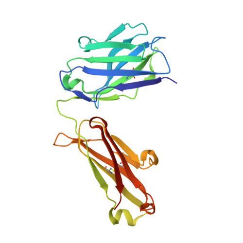
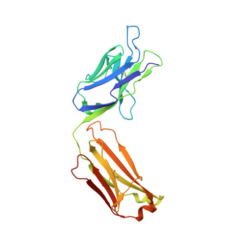
MoFvAb: Modeling the Fv region of antibodies.

Bujotzek, A.Fuchs, A.Qu, C.Benz, J.Klostermann, S.Antes, I.Georges, G.

(2015) MAbs 7: 838-852

  • DOI: https://doi.org/10.1080/19420862.2015.1068492
  • Primary Citation of Related Structures:  
    5BMF

  • PubMed Abstract: 

    Knowledge of the 3-dimensional structure of the antigen-binding region of antibodies enables numerous useful applications regarding the design and development of antibody-based drugs. We present a knowledge-based antibody structure prediction methodology that incorporates concepts that have arisen from an applied antibody engineering environment. The protocol exploits the rich and continuously growing supply of experimentally derived antibody structures available to predict CDR loop conformations and the packing of heavy and light chain quickly and without user intervention. The homology models are refined by a novel antibody-specific approach to adapt and rearrange sidechains based on their chemical environment. The method achieves very competitive all-atom root mean square deviation values in the order of 1.5 Å on different evaluation datasets consisting of both known and previously unpublished antibody crystal structures.


  • Organizational Affiliation

    a Roche Pharmaceutical Research and Early Development; Large Molecule Research; Roche Innovation Center Penzberg ; Penzberg , Germany.


Macromolecules
Find similar proteins by:  (by identity cutoff)  |  3D Structure
Entity ID: 1
MoleculeChains Sequence LengthOrganismDetailsImage
Fab fragment heavy chainA [auth H]224Homo sapiensMutation(s): 0 
Entity Groups  
Sequence Clusters30% Identity50% Identity70% Identity90% Identity95% Identity100% Identity
Sequence Annotations
Expand
  • Reference Sequence
Find similar proteins by:  (by identity cutoff)  |  3D Structure
Entity ID: 2
MoleculeChains Sequence LengthOrganismDetailsImage
Fab fragment light chainB [auth L]219Homo sapiensMutation(s): 0 
Entity Groups  
Sequence Clusters30% Identity50% Identity70% Identity90% Identity95% Identity100% Identity
Sequence Annotations
Expand
  • Reference Sequence
Small Molecules
Ligands 1 Unique
IDChains Name / Formula / InChI Key2D Diagram3D Interactions
4TJ
Query on 4TJ

Download Ideal Coordinates CCD File 
C [auth H]2-(5-{1-[1-(1,3-dimethyl-2,6-dioxo-2,3,6,7-tetrahydro-1H-purin-8-yl)-4,15-dioxo-8,11-dioxa-5,14-diazaicosan-20-yl]-3,3-dimethyl-6-sulfo-1,3-dihydro-2H-indol-2-ylidene}penta-1,3-dien-1-yl)-1-ethyl-3,3-dimethyl-3H-indolium-5-sulfonate
C50 H66 N8 O12 S2
PHMVJTFYMDAUCS-UHFFFAOYSA-N
Experimental Data & Validation

Experimental Data

  • Method: X-RAY DIFFRACTION
  • Resolution: 2.80 Å
  • R-Value Free: 0.220 
  • R-Value Work: 0.186 
  • R-Value Observed: 0.188 
  • Space Group: P 31 2 1
Unit Cell:
Length ( Å )Angle ( ˚ )
a = 129.438α = 90
b = 129.438β = 90
c = 92.692γ = 120
Software Package:
Software NamePurpose
PHENIXrefinement
SCALAdata scaling
PHASERphasing
MOSFLMdata reduction

Structure Validation

View Full Validation Report


Ligand Structure Quality Assessment 


Entry History 

Deposition Data

Revision History  (Full details and data files)

  • Version 1.0: 2015-07-29
    Type: Initial release
  • Version 1.1: 2015-09-02
    Changes: Database references