5ANM

Crystal structure of IgE Fc in complex with a neutralizing antibody


Experimental Data Snapshot

  • Method: X-RAY DIFFRACTION
  • Resolution: 2.85 Å
  • R-Value Free: 0.260 
  • R-Value Work: 0.196 
  • R-Value Observed: 0.199 

wwPDB Validation   3D Report Full Report


This is version 2.1 of the entry. See complete history


Literature

A Novel Ige-Neutralizing Antibody for the Treatment of Severe Uncontrolled Asthma.

Cohen, E.S.Dobson, C.L.Kack, H.Wang, B.Sims, D.A.Lloyd, C.O.England, E.Rees, D.G.Guo, H.Karagiannis, S.N.O'Brien, S.Persdotter, S.Ekdahl, H.Butler, R.Keyes, F.Oakley, S.Carlsson, M.Briend, E.Wilkinson, T.Anderson, I.K.Monk, P.D.Von Wachenfeldt, K.Eriksson, P.F.Gould, H.J.Vaughan, T.J.May, R.D.

(2015) MAbs 6: 756

  • DOI: https://doi.org/10.4161/mabs.28394
  • Primary Citation of Related Structures:  
    5ANM

  • PubMed Abstract: 

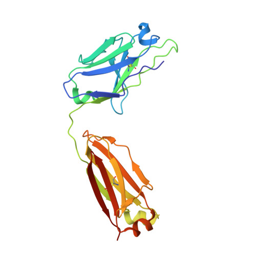
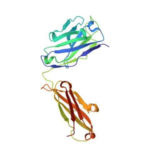
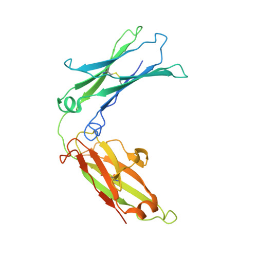
    The critical role played by IgE in allergic asthma is well-documented and clinically precedented, but some patients in whom IgE neutralization may still offer clinical benefit are excluded from treatment with the existing anti-IgE therapy, omalizumab, due to high total IgE levels or body mass. In this study, we sought to generate a novel high affinity anti-IgE antibody (MEDI4212) with potential to treat a broad severe asthma patient population. Analysis of body mass, total and allergen-specific IgE levels in a cohort of severe asthmatics was used to support the rationale for development of a high affinity IgE-targeted antibody therapeutic. Phage display technology was used to generate a human IgG1 lead antibody, MEDI4212, which was characterized in vitro using binding, signaling and functional assay systems. Protein crystallography was used to determine the details of the interaction between MEDI4212 and IgE. MEDI4212 bound human IgE with an affinity of 1.95 pM and was shown to target critical residues in the IgE Cε3 domain critical for interaction with FcεRI. MEDI4212 potently inhibited responses through FcεRI and also prevented the binding of IgE to CD23. When used ex vivo at identical concentration, MEDI4212 depleted free-IgE from human sera to levels ~1 log lower than omalizumab. Our results thus indicate that MEDI4212 is a novel, high affinity antibody that binds specifically to IgE and prevents IgE binding to its receptors. MEDI4212 effectively depleted free-IgE from human sera ex vivo to a level (1 IU/mL) anticipated to provide optimal IgE suppression in severe asthma patients.


  • Organizational Affiliation

    MedImmune Ltd; Cambridge, UK.


Macromolecules
Find similar proteins by:  (by identity cutoff)  |  3D Structure
Entity ID: 1
MoleculeChains Sequence LengthOrganismDetailsImage
IMMUNOGLOBULIN GA,
C,
I [auth L]
217Homo sapiensMutation(s): 0 
Entity Groups  
Sequence Clusters30% Identity50% Identity70% Identity90% Identity95% Identity100% Identity
Sequence Annotations
Expand
  • Reference Sequence
Find similar proteins by:  (by identity cutoff)  |  3D Structure
Entity ID: 2
MoleculeChains Sequence LengthOrganismDetailsImage
IMMUNOGLOBULIN G
B, D, H
229Homo sapiensMutation(s): 0 
Entity Groups  
Sequence Clusters30% Identity50% Identity70% Identity90% Identity95% Identity100% Identity
Sequence Annotations
Expand
  • Reference Sequence
Find similar proteins by:  (by identity cutoff)  |  3D Structure
Entity ID: 3
MoleculeChains Sequence LengthOrganismDetailsImage
IG EPSILON CHAIN C REGION
E, F, G
242Homo sapiensMutation(s): 0 
UniProt & NIH Common Fund Data Resources
Find proteins for P01854 (Homo sapiens)
Explore P01854 
Go to UniProtKB:  P01854
PHAROS:  P01854
GTEx:  ENSG00000211891 
Entity Groups  
Sequence Clusters30% Identity50% Identity70% Identity90% Identity95% Identity100% Identity
UniProt GroupP01854
Sequence Annotations
Expand
  • Reference Sequence
Oligosaccharides

Help

Entity ID: 4
MoleculeChains Length2D Diagram Glycosylation3D Interactions
alpha-D-mannopyranose-(1-3)-alpha-D-mannopyranose-(1-6)-[alpha-D-mannopyranose-(1-3)]beta-D-mannopyranose-(1-4)-2-acetamido-2-deoxy-beta-D-glucopyranose-(1-4)-2-acetamido-2-deoxy-beta-D-glucopyranoseJ [auth I]6N-Glycosylation
Glycosylation Resources
GlyTouCan:  G09724ZC
GlyCosmos:  G09724ZC
GlyGen:  G09724ZC
Entity ID: 5
MoleculeChains Length2D Diagram Glycosylation3D Interactions
beta-D-mannopyranose-(1-3)-[alpha-D-mannopyranose-(1-6)]beta-D-mannopyranose-(1-4)-2-acetamido-2-deoxy-beta-D-glucopyranose-(1-4)-2-acetamido-2-deoxy-beta-D-glucopyranoseK [auth J]5N-Glycosylation
Glycosylation Resources
GlyTouCan:  G42769XG
GlyCosmos:  G42769XG
GlyGen:  G42769XG
Entity ID: 6
MoleculeChains Length2D Diagram Glycosylation3D Interactions
alpha-D-mannopyranose-(1-3)-alpha-D-mannopyranose-(1-6)-[beta-D-mannopyranose-(1-3)]beta-D-mannopyranose-(1-4)-2-acetamido-2-deoxy-beta-D-glucopyranose-(1-4)-2-acetamido-2-deoxy-beta-D-glucopyranoseL [auth K]6N-Glycosylation
Glycosylation Resources
GlyTouCan:  G00601VC
GlyCosmos:  G00601VC
GlyGen:  G00601VC
Experimental Data & Validation

Experimental Data

  • Method: X-RAY DIFFRACTION
  • Resolution: 2.85 Å
  • R-Value Free: 0.260 
  • R-Value Work: 0.196 
  • R-Value Observed: 0.199 
  • Space Group: P 32 2 1
Unit Cell:
Length ( Å )Angle ( ˚ )
a = 140.957α = 90
b = 140.957β = 90
c = 244.448γ = 120
Software Package:
Software NamePurpose
REFMACrefinement
MOSFLMdata reduction
SCALAdata scaling
PHASERphasing

Structure Validation

View Full Validation Report


Entry History 

Revision History  (Full details and data files)

  • Version 1.0: 2015-09-30
    Type: Initial release
  • Version 2.0: 2020-07-29
    Type: Remediation
    Reason: Carbohydrate remediation
    Changes: Advisory, Atomic model, Data collection, Derived calculations, Other, Structure summary
  • Version 2.1: 2024-01-10
    Changes: Data collection, Database references, Refinement description, Structure summary