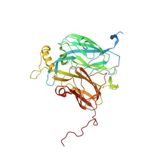
Sub-Atomic Resolution X-Ray Crystallography and Neutron Crystallography: Promise, Challenges and Potential.
Blakeley, M.P., Hasnain, S.S., Antonyuk, S.V.(2015) IUCrJ 2: 464
- PubMed: 26175905
- DOI: https://doi.org/10.1107/S2052252515011239
- Primary Citation of Related Structures:
5AKR - PubMed Abstract:
The International Year of Crystallography saw the number of macromolecular structures deposited in the Protein Data Bank cross the 100000 mark, with more than 90000 of these provided by X-ray crystallography. The number of X-ray structures determined to sub-atomic resolution (i.e. ≤1 Å) has passed 600 and this is likely to continue to grow rapidly with diffraction-limited synchrotron radiation sources such as MAX-IV (Sweden) and Sirius (Brazil) under construction. A dozen X-ray structures have been deposited to ultra-high resolution (i.e. ≤0.7 Å), for which precise electron density can be exploited to obtain charge density and provide information on the bonding character of catalytic or electron transfer sites. Although the development of neutron macromolecular crystallography over the years has been far less pronounced, and its application much less widespread, the availability of new and improved instrumentation, combined with dedicated deuteration facilities, are beginning to transform the field. Of the 83 macromolecular structures deposited with neutron diffraction data, more than half (49/83, 59%) were released since 2010. Sub-mm(3) crystals are now regularly being used for data collection, structures have been determined to atomic resolution for a few small proteins, and much larger unit-cell systems (cell edges >100 Å) are being successfully studied. While some details relating to H-atom positions are tractable with X-ray crystallography at sub-atomic resolution, the mobility of certain H atoms precludes them from being located. In addition, highly polarized H atoms and protons (H(+)) remain invisible with X-rays. Moreover, the majority of X-ray structures are determined from cryo-cooled crystals at 100 K, and, although radiation damage can be strongly controlled, especially since the advent of shutterless fast detectors, and by using limited doses and crystal translation at micro-focus beams, radiation damage can still take place. Neutron crystallography therefore remains the only approach where diffraction data can be collected at room temperature without radiation damage issues and the only approach to locate mobile or highly polarized H atoms and protons. Here a review of the current status of sub-atomic X-ray and neutron macromolecular crystallography is given and future prospects for combined approaches are outlined. New results from two metalloproteins, copper nitrite reductase and cytochrome c', are also included, which illustrate the type of information that can be obtained from sub-atomic-resolution (∼0.8 Å) X-ray structures, while also highlighting the need for complementary neutron studies that can provide details of H atoms not provided by X-ray crystallography.
Organizational Affiliation:
Large-Scale Structures Group, Institut Laue-Langevin , 71 Avenue des Martyrs, Grenoble 38000, France.


