5AK9

THE CRYSTAL STRUCTURE OF I-DMOI Q42AK120M IN COMPLEX WITH ITS TARGET DNA IN THE PRESENCE OF 2MM MN


Experimental Data Snapshot

  • Method: X-RAY DIFFRACTION
  • Resolution: 2.60 Å
  • R-Value Free: 0.222 
  • R-Value Work: 0.170 
  • R-Value Observed: 0.172 

wwPDB Validation   3D Report Full Report


This is version 1.3 of the entry. See complete history


Literature

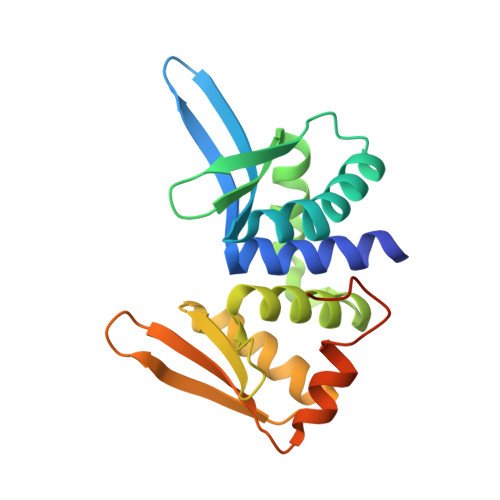
Engineering a Nickase on the Homing Endonuclease I-Dmoi Scaffold.

Molina, R.Marcaida, M.J.Redondo, P.Marenchino, M.Duchateau, P.D'Abramo, M.Montoya, G.Prieto, J.

(2015) J Biol Chem 290: 18534

  • DOI: https://doi.org/10.1074/jbc.M115.658666
  • Primary Citation of Related Structures:  
    5AK9, 5AKF, 5AKM, 5AKN

  • PubMed Abstract: 

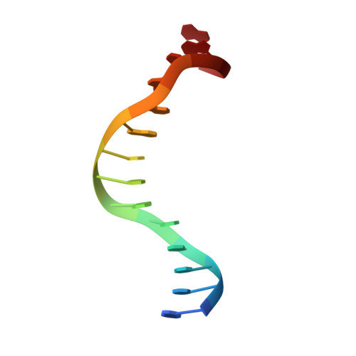
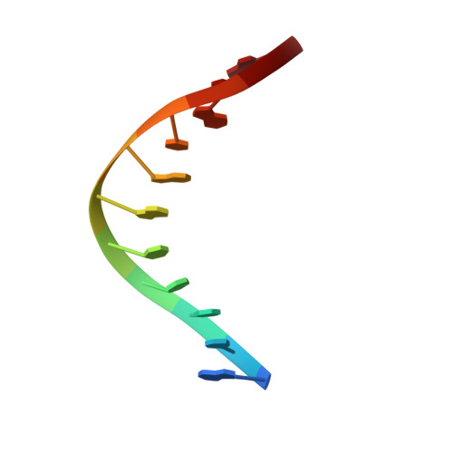
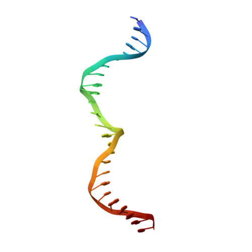
    Homing endonucleases are useful tools for genome modification because of their capability to recognize and cleave specifically large DNA targets. These endonucleases generate a DNA double strand break that can be repaired by the DNA damage response machinery. The break can be repaired by homologous recombination, an error-free mechanism, or by non-homologous end joining, a process susceptible to introducing errors in the repaired sequence. The type of DNA cleavage might alter the balance between these two alternatives. The use of "nickases" producing a specific single strand break instead of a double strand break could be an approach to reduce the toxicity associated with non-homologous end joining by promoting the use of homologous recombination to repair the cleavage of a single DNA break. Taking advantage of the sequential DNA cleavage mechanism of I-DmoI LAGLIDADG homing endonuclease, we have developed a new variant that is able to cut preferentially the coding DNA strand, generating a nicked DNA target. Our structural and biochemical analysis shows that by decoupling the action of the catalytic residues acting on each strand we can inhibit one of them while keeping the other functional.


  • Organizational Affiliation

    From the Macromolecular Crystallography Group and.


Macromolecules

Find similar proteins by:  (by identity cutoff)  |  3D Structure
Entity ID: 1
MoleculeChains Sequence LengthOrganismDetailsImage
HOMING ENDONUCLEASE I-DMOIA,
E,
I [auth J]
199Desulfurococcus mucosusMutation(s): 0 
EC: 3.1
UniProt
Find proteins for P21505 (Desulfurococcus mucosus)
Explore P21505 
Go to UniProtKB:  P21505
Entity Groups  
Sequence Clusters30% Identity50% Identity70% Identity90% Identity95% Identity100% Identity
UniProt GroupP21505
Sequence Annotations
Expand
  • Reference Sequence

Find similar nucleic acids by:  Sequence   |   3D Structure  

Entity ID: 2
MoleculeChains LengthOrganismImage
5'-D(*GP*CP*CP*TP*TP*GP*CP*CP*GP*GP*GP*TP*AP*AP)-3'B,
F,
J [auth K]
14synthetic construct
Sequence Annotations
Expand
  • Reference Sequence

Find similar nucleic acids by:  Sequence   |   3D Structure  

Entity ID: 3
MoleculeChains LengthOrganismImage
5'-D(*GP*TP*TP*CP*CP*GP*GP*CP*GP*CP*GP)-3C,
G,
K [auth L]
11synthetic construct
Sequence Annotations
Expand
  • Reference Sequence

Find similar nucleic acids by:  Sequence   |   3D Structure  

Entity ID: 4
MoleculeChains LengthOrganismImage
25MERD,
H,
L [auth N]
25synthetic construct
Sequence Annotations
Expand
  • Reference Sequence
Experimental Data & Validation

Experimental Data

  • Method: X-RAY DIFFRACTION
  • Resolution: 2.60 Å
  • R-Value Free: 0.222 
  • R-Value Work: 0.170 
  • R-Value Observed: 0.172 
  • Space Group: P 1 21 1
Unit Cell:
Length ( Å )Angle ( ˚ )
a = 107.53α = 90
b = 70.88β = 119.78
c = 107.68γ = 90
Software Package:
Software NamePurpose
PHENIXrefinement
XDSdata reduction
SCALAdata scaling
PHASERphasing

Structure Validation

View Full Validation Report


Entry History 

Deposition Data

Revision History  (Full details and data files)

  • Version 1.0: 2015-06-17
    Type: Initial release
  • Version 1.1: 2015-08-05
    Changes: Database references
  • Version 1.2: 2017-08-23
    Changes: Data collection
  • Version 1.3: 2024-01-10
    Changes: Data collection, Database references, Derived calculations, Other, Refinement description