5ADO

Crystal structure of the paraoxon-modified A.17 antibody FAB fragment - Light chain S35R mutant


Experimental Data Snapshot

  • Method: X-RAY DIFFRACTION
  • Resolution: 1.55 Å
  • R-Value Free: 0.199 
  • R-Value Work: 0.168 
  • R-Value Observed: 0.170 

wwPDB Validation   3D Report Full Report


This is version 1.2 of the entry. See complete history


Literature

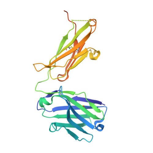
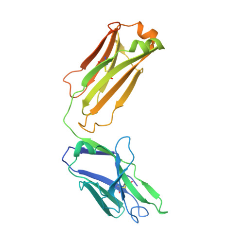
Robotic Qm/Mm-Driven Maturation of Antibody Combining Sites.

Smirnov, I.V.Golovin, A.V.Chatziefthimiou, S.D.Stepanova, A.V.Peng, Y.Zolotareva, O.I.Belogurov, A.A.Kurkova, I.N.Ponomarenko, N.A.Wilmanns, M.Blackburn, G.M.Gabibov, A.G.Lerner, R.A.

(2016) Sci Adv 2: 01695

  • DOI: https://doi.org/10.1126/sciadv.1501695
  • Primary Citation of Related Structures:  
    5ADO, 5ADP

  • PubMed Abstract: 

    In vitro selection of antibodies from large repertoires of immunoglobulin (Ig) combining sites using combinatorial libraries is a powerful tool, with great potential for generating in vivo scavengers for toxins. However, addition of a maturation function is necessary to enable these selected antibodies to more closely mimic the full mammalian immune response. We approached this goal using quantum mechanics/molecular mechanics (QM/MM) calculations to achieve maturation in silico. We preselected A17, an Ig template, from a naïve library for its ability to disarm a toxic pesticide related to organophosphorus nerve agents. Virtual screening of 167,538 robotically generated mutants identified an optimum single point mutation, which experimentally boosted wild-type Ig scavenger performance by 170-fold. We validated the QM/MM predictions via kinetic analysis and crystal structures of mutant apo-A17 and covalently modified Ig, thereby identifying the displacement of one water molecule by an arginine as delivering this catalysis.


  • Organizational Affiliation

    Shemyakin-Ovchinnikov Institute of Bioorganic Chemistry, Russian Academy of Sciences, ulitsa Miklukho-Maklaya 16/10, 117997 Moscow V-437, Russian Federation.


Macromolecules
Find similar proteins by:  (by identity cutoff)  |  3D Structure
Entity ID: 1
MoleculeChains Sequence LengthOrganismDetailsImage
FAB A.17A [auth H]255Homo sapiensMutation(s): 0 
Entity Groups  
Sequence Clusters30% Identity50% Identity70% Identity90% Identity95% Identity100% Identity
Sequence Annotations
Expand
  • Reference Sequence
Find similar proteins by:  (by identity cutoff)  |  3D Structure
Entity ID: 2
MoleculeChains Sequence LengthOrganismDetailsImage
FAB A.17B [auth L]247Homo sapiensMutation(s): 0 
Entity Groups  
Sequence Clusters30% Identity50% Identity70% Identity90% Identity95% Identity100% Identity
Sequence Annotations
Expand
  • Reference Sequence
Small Molecules
Ligands 1 Unique
IDChains Name / Formula / InChI Key2D Diagram3D Interactions
DEP
Query on DEP

Download Ideal Coordinates CCD File 
C [auth L]DIETHYL PHOSPHONATE
C4 H11 O3 P
MJUJXFBTEFXVKU-UHFFFAOYSA-N
Experimental Data & Validation

Experimental Data

  • Method: X-RAY DIFFRACTION
  • Resolution: 1.55 Å
  • R-Value Free: 0.199 
  • R-Value Work: 0.168 
  • R-Value Observed: 0.170 
  • Space Group: P 1 21 1
Unit Cell:
Length ( Å )Angle ( ˚ )
a = 52.661α = 90
b = 67.075β = 107.52
c = 66.698γ = 90
Software Package:
Software NamePurpose
PHENIXrefinement
XDSdata reduction
SCALAdata scaling
MOLREPphasing

Structure Validation

View Full Validation Report


Entry History 

Revision History  (Full details and data files)

  • Version 1.0: 2016-11-09
    Type: Initial release
  • Version 1.1: 2018-03-07
    Changes: Data collection, Source and taxonomy
  • Version 1.2: 2024-01-10
    Changes: Data collection, Database references, Derived calculations, Other, Refinement description