5A8M

Crystal structure of the selenomethionine derivative of beta-glucanase SdGluc5_26A from Saccharophagus degradans


Experimental Data Snapshot

  • Method: X-RAY DIFFRACTION
  • Resolution: 1.86 Å
  • R-Value Free: 0.165 
  • R-Value Work: 0.143 
  • R-Value Observed: 0.144 

wwPDB Validation   3D Report Full Report


This is version 1.2 of the entry. See complete history


Literature

The Quaternary Structure of a Glycoside Hydrolase Dictates Specificity Towards Beta-Glucans

Lafond, M.Sulzenbacher, G.Freyd, T.Henrissat, B.Berrin, J.G.Garron, M.L.

(2016) J Biol Chem 291: 7183

  • DOI: https://doi.org/10.1074/jbc.M115.695999
  • Primary Citation of Related Structures:  
    5A8M, 5A8N

  • PubMed Abstract: 

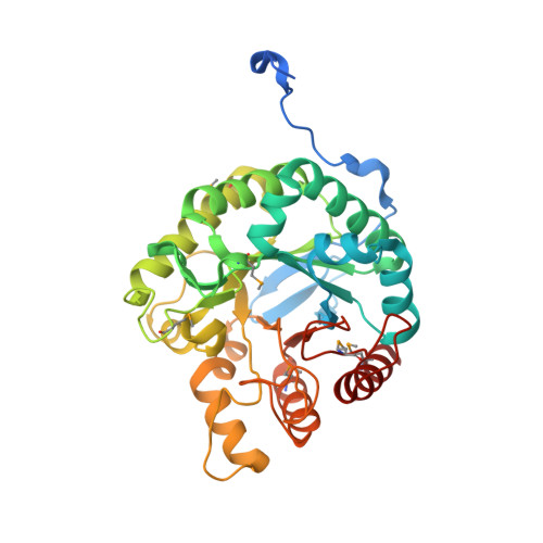
    In the Carbohydrate-Active Enzyme (CAZy) database, glycoside hydrolase family 5 (GH5) is a large family with more than 6,000 sequences. Among the 51 described GH5 subfamilies, subfamily GH5_26 contains members that display either endo-β(1,4)-glucanase or β(1,3;1,4)-glucanase activities. In this study, we focused on the GH5_26 enzyme fromSaccharophagus degradans(SdGluc5_26A), a marine bacterium known for its capacity to degrade a wide diversity of complex polysaccharides.SdGluc5_26A displays lichenase activity toward β(1,3;1,4)-glucans with a side cellobiohydrolase activity toward β(1,4)-glucans. The three-dimensional structure ofSdGluc5_26A adopts a stable trimeric quaternary structure also observable in solution. The N-terminal region ofSdGluc5_26A protrudes into the active site of an adjacent monomer. To understand whether this occupation of the active site could influence its activity, we conducted a comprehensive enzymatic characterization ofSdGluc5_26A and of a mutant truncated at the N terminus. Ligand complex structures and kinetic analyses reveal that the N terminus governs the substrate specificity ofSdGluc5_26A. Its deletion opens the enzyme cleft at the -3 subsite and turns the enzyme into an endo-β(1,4)-glucanase. This study demonstrates that experimental approaches can reveal structure-function relationships out of reach of current bioinformatic predictions.


  • Organizational Affiliation

    From the Institut des Sciences Moléculaires de Marseille-BiosCiences, UMR7313 CNRS, Aix-Marseille University, Pôle de l'Etoile, 13284 Marseille, France, the INRA, UMR1163, Biodiversité et Biotechnologie Fongiques, Aix-Marseille University, Polytech'Marseille, F-13288 Marseille, France.


Macromolecules
Find similar proteins by:  (by identity cutoff)  |  3D Structure
Entity ID: 1
MoleculeChains Sequence LengthOrganismDetailsImage
PUTATIVE RETAINING B-GLYCOSIDASE
A, B, C
365Saccharophagus degradans 2-40Mutation(s): 0 
EC: 3.2.1.73
UniProt
Find proteins for Q21KE5 (Saccharophagus degradans (strain 2-40 / ATCC 43961 / DSM 17024))
Explore Q21KE5 
Go to UniProtKB:  Q21KE5
Entity Groups  
Sequence Clusters30% Identity50% Identity70% Identity90% Identity95% Identity100% Identity
UniProt GroupQ21KE5
Sequence Annotations
Expand
  • Reference Sequence
Small Molecules
Ligands 5 Unique
IDChains Name / Formula / InChI Key2D Diagram3D Interactions
PGE
Query on PGE

Download Ideal Coordinates CCD File 
AA [auth B]
BA [auth B]
CA [auth B]
DA [auth B]
EA [auth B]
AA [auth B],
BA [auth B],
CA [auth B],
DA [auth B],
EA [auth B],
FA [auth B],
GA [auth B],
HA [auth B],
L [auth A],
M [auth A],
N [auth A],
O [auth A],
P [auth A],
PA [auth C],
Q [auth A],
QA [auth C],
R [auth A],
RA [auth C],
SA [auth C],
TA [auth C],
UA [auth C],
Z [auth B]
TRIETHYLENE GLYCOL
C6 H14 O4
ZIBGPFATKBEMQZ-UHFFFAOYSA-N
SO4
Query on SO4

Download Ideal Coordinates CCD File 
H [auth A],
MA [auth C],
W [auth B]
SULFATE ION
O4 S
QAOWNCQODCNURD-UHFFFAOYSA-L
GOL
Query on GOL

Download Ideal Coordinates CCD File 
I [auth A]
J [auth A]
K [auth A]
NA [auth C]
OA [auth C]
I [auth A],
J [auth A],
K [auth A],
NA [auth C],
OA [auth C],
X [auth B],
Y [auth B]
GLYCEROL
C3 H8 O3
PEDCQBHIVMGVHV-UHFFFAOYSA-N
CL
Query on CL

Download Ideal Coordinates CCD File 
D [auth A]
E [auth A]
F [auth A]
IA [auth C]
JA [auth C]
D [auth A],
E [auth A],
F [auth A],
IA [auth C],
JA [auth C],
KA [auth C],
S [auth B],
T [auth B],
U [auth B]
CHLORIDE ION
Cl
VEXZGXHMUGYJMC-UHFFFAOYSA-M
MG
Query on MG

Download Ideal Coordinates CCD File 
G [auth A],
LA [auth C],
V [auth B]
MAGNESIUM ION
Mg
JLVVSXFLKOJNIY-UHFFFAOYSA-N
Modified Residues  1 Unique
IDChains TypeFormula2D DiagramParent
MSE
Query on MSE
A, B, C
L-PEPTIDE LINKINGC5 H11 N O2 SeMET
Experimental Data & Validation

Experimental Data

  • Method: X-RAY DIFFRACTION
  • Resolution: 1.86 Å
  • R-Value Free: 0.165 
  • R-Value Work: 0.143 
  • R-Value Observed: 0.144 
  • Space Group: P 41 21 2
Unit Cell:
Length ( Å )Angle ( ˚ )
a = 142.605α = 90
b = 142.605β = 90
c = 136.277γ = 90
Software Package:
Software NamePurpose
REFMACrefinement
XDSdata reduction
SCALAdata scaling
SHELXphasing

Structure Validation

View Full Validation Report


Entry History 

Deposition Data

Revision History  (Full details and data files)

  • Version 1.0: 2016-01-20
    Type: Initial release
  • Version 1.1: 2016-04-06
    Changes: Database references
  • Version 1.2: 2017-09-13
    Changes: Data collection, Structure summary