4ZV8

Structure of CYP2B6 (Y226H/K262R) with additional mutation Y244W in complex with alpha-Pinene


Experimental Data Snapshot

  • Method: X-RAY DIFFRACTION
  • Resolution: 2.24 Å
  • R-Value Free: 0.232 
  • R-Value Work: 0.169 
  • R-Value Observed: 0.172 

wwPDB Validation   3D Report Full Report


Ligand Structure Quality Assessment 


This is version 1.5 of the entry. See complete history


Literature

Coumarin Derivatives as Substrate Probes of Mammalian Cytochromes P450 2B4 and 2B6: Assessing the Importance of 7-Alkoxy Chain Length, Halogen Substitution, and Non-Active Site Mutations.

Liu, J.Shah, M.B.Zhang, Q.Stout, C.D.Halpert, J.R.Wilderman, P.R.

(2016) Biochemistry 55: 1997-2007

  • DOI: https://doi.org/10.1021/acs.biochem.5b01330
  • Primary Citation of Related Structures:  
    4ZV8, 5EM4

  • PubMed Abstract: 

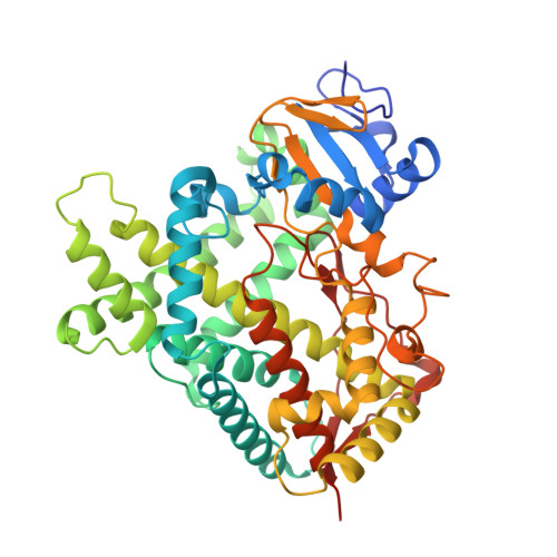
    Using a combined structural and biochemical approach, the functional importance of a recently described peripheral pocket bounded by the E-, F-, G-, and I-helices in CYP2B4 and 2B6 was probed. Three series of 4-substituted-7-alkoxycoumarin derivatives with -H, -CH3, or -CF3 at the 4 position of the coumarin core were used initially to monitor functional differences between CYP2B4 and 2B6. 7-Ethoxy-4-(trifluoromethyl)coumarin (7-EFC) displayed the highest catalytic efficiency among these substrates. Mutants were made to alter side-chain polarity (V/E194Q) or bulk (F/Y244W) to alter access to the peripheral pocket. Modest increases in catalytic efficiency of 7-EFC O-deethylation by the mutants were magnified considerably by chlorination or bromination of the substrate ethoxy chain. A structure of CYP2B6 Y244W in complex with (+)-α-pinene was solved at 2.2 Å and showed no CYMAL-5 in the peripheral pocket. A ligand free structure of CYP2B4 F244W was solved at 3.0 Å with CYMAL-5 in the peripheral pocket. In both instances, comparison of the respective wild-type and mutant CYP2B enzymes revealed that CYMAL-5 occupancy of the peripheral pocket had little effect on the topology of active site residue side-chains, despite the fact that the peripheral pocket and active site are located on opposite sides of the I-helix. Analysis of available CYP2B structures suggest that the effect of the amino acid substitutions within the peripheral pocket derive from altered interactions between the F and G helices.


  • Organizational Affiliation

    School of Pharmacy, University of Connecticut , Storrs, Connecticut 06269, United States.


Macromolecules
Find similar proteins by:  (by identity cutoff)  |  3D Structure
Entity ID: 1
MoleculeChains Sequence LengthOrganismDetailsImage
Cytochrome P450 2B6476Homo sapiensMutation(s): 3 
Gene Names: CYP2B6
EC: 1.14.13
Membrane Entity: Yes 
UniProt & NIH Common Fund Data Resources
Find proteins for P20813 (Homo sapiens)
Explore P20813 
Go to UniProtKB:  P20813
PHAROS:  P20813
GTEx:  ENSG00000197408 
Entity Groups  
Sequence Clusters30% Identity50% Identity70% Identity90% Identity95% Identity100% Identity
UniProt GroupP20813
Sequence Annotations
Expand
  • Reference Sequence
Experimental Data & Validation

Experimental Data

  • Method: X-RAY DIFFRACTION
  • Resolution: 2.24 Å
  • R-Value Free: 0.232 
  • R-Value Work: 0.169 
  • R-Value Observed: 0.172 
  • Space Group: P 32 2 1
Unit Cell:
Length ( Å )Angle ( ˚ )
a = 77.24α = 90
b = 77.24β = 90
c = 204.25γ = 120
Software Package:
Software NamePurpose
REFMACrefinement
MOSFLMdata reduction
SCALAdata scaling
Cootmodel building

Structure Validation

View Full Validation Report


Ligand Structure Quality Assessment 


Entry History & Funding Information

Deposition Data


Funding OrganizationLocationGrant Number
National Institutes of Health/National Institute of General Medical Sciences (NIH/NIGMS)United StatesGM098538

Revision History  (Full details and data files)

  • Version 1.0: 2016-03-30
    Type: Initial release
  • Version 1.1: 2016-07-13
    Changes: Database references
  • Version 1.2: 2017-09-06
    Changes: Author supporting evidence, Database references, Derived calculations
  • Version 1.3: 2017-11-22
    Changes: Refinement description
  • Version 1.4: 2019-12-25
    Changes: Author supporting evidence
  • Version 1.5: 2023-09-27
    Changes: Data collection, Database references, Refinement description