4ZUO

Crystal structure of acetylpolyamine amidohydrolase from Mycoplana ramosa in complex with a hydroxamate inhibitor


Experimental Data Snapshot

  • Method: X-RAY DIFFRACTION
  • Resolution: 1.33 Å
  • R-Value Free: 0.147 
  • R-Value Work: 0.133 
  • R-Value Observed: 0.134 

wwPDB Validation   3D Report Full Report


Ligand Structure Quality Assessment 


This is version 1.5 of the entry. See complete history


Literature

Design, Synthesis, and Evaluation of Polyamine Deacetylase Inhibitors, and High-Resolution Crystal Structures of Their Complexes with Acetylpolyamine Amidohydrolase.

Decroos, C.Christianson, D.W.

(2015) Biochemistry 54: 4692-4703

  • DOI: https://doi.org/10.1021/acs.biochem.5b00536
  • Primary Citation of Related Structures:  
    4ZUM, 4ZUN, 4ZUO, 4ZUP, 4ZUQ, 4ZUR

  • PubMed Abstract: 

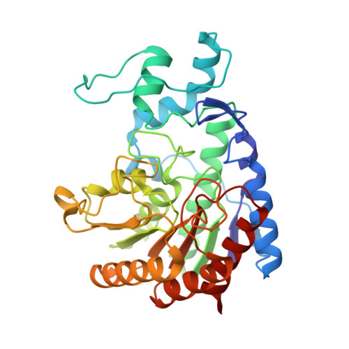
    Polyamines are essential aliphatic polycations that bind to nucleic acids and accordingly are involved in a variety of cellular processes. Polyamine function can be regulated by acetylation and deacetylation, just as histone function can be regulated by lysine acetylation and deacetylation. Acetylpolyamine amidohydrolase (APAH) from Mycoplana ramosa is a zinc-dependent polyamine deacetylase that shares approximately 20% amino acid sequence identity with human histone deacetylases. We now report the X-ray crystal structures of APAH-inhibitor complexes in a new and superior crystal form that diffracts to very high resolution (1.1-1.4 Å). Inhibitors include previously synthesized analogues of N(8)-acetylspermidine bearing trifluoromethylketone, thiol, and hydroxamate zinc-binding groups [Decroos, C., Bowman, C. M., and Christianson, D. W. (2013) Bioorg. Med. Chem. 21, 4530], and newly synthesized hydroxamate analogues of shorter, monoacetylated diamines, the most potent of which is the hydroxamate analogue of N-acetylcadaverine (IC50 = 68 nM). The high-resolution crystal structures of APAH-inhibitor complexes provide key inferences about the inhibition and catalytic mechanism of zinc-dependent deacetylases. For example, the trifluoromethylketone analogue of N(8)-acetylspermidine binds as a tetrahedral gem-diol that mimics the tetrahedral intermediate and its flanking transition states in catalysis. Surprisingly, this compound is also a potent inhibitor of human histone deacetylase 8 with an IC50 of 260 nM. Crystal structures of APAH-inhibitor complexes are determined at the highest resolution of any currently existing zinc deacetylase structure and thus represent the most accurate reference points for understanding structure-mechanism and structure-inhibition relationships in this critically important enzyme family.


  • Organizational Affiliation

    Roy and Diana Vagelos Laboratories, Department of Chemistry, University of Pennsylvania, Philadelphia, Pennsylvania 19104-6323, United States.


Macromolecules
Find similar proteins by:  (by identity cutoff)  |  3D Structure
Entity ID: 1
MoleculeChains Sequence LengthOrganismDetailsImage
Acetylpolyamine aminohydrolase
A, B
341Mycoplana ramosaMutation(s): 0 
Gene Names: aphAaph
UniProt
Find proteins for Q48935 (Mycoplana ramosa)
Explore Q48935 
Go to UniProtKB:  Q48935
Entity Groups  
Sequence Clusters30% Identity50% Identity70% Identity90% Identity95% Identity100% Identity
UniProt GroupQ48935
Sequence Annotations
Expand
  • Reference Sequence
Small Molecules
Ligands 5 Unique
IDChains Name / Formula / InChI Key2D Diagram3D Interactions
XS6
Query on XS6

Download Ideal Coordinates CCD File 
F [auth A],
L [auth B]
6-[(3-aminopropyl)amino]-N-hydroxyhexanamide
C9 H21 N3 O2
BREASWSBOVLQOH-UHFFFAOYSA-N
GOL
Query on GOL

Download Ideal Coordinates CCD File 
G [auth A],
H [auth A],
M [auth B],
N [auth B],
O [auth B]
GLYCEROL
C3 H8 O3
PEDCQBHIVMGVHV-UHFFFAOYSA-N
ZN
Query on ZN

Download Ideal Coordinates CCD File 
C [auth A],
I [auth B]
ZINC ION
Zn
PTFCDOFLOPIGGS-UHFFFAOYSA-N
K
Query on K

Download Ideal Coordinates CCD File 
D [auth A],
J [auth B]
POTASSIUM ION
K
NPYPAHLBTDXSSS-UHFFFAOYSA-N
NH4
Query on NH4

Download Ideal Coordinates CCD File 
E [auth A],
K [auth B]
AMMONIUM ION
H4 N
QGZKDVFQNNGYKY-UHFFFAOYSA-O
Binding Affinity Annotations 
IDSourceBinding Affinity
XS6 BindingDB:  4ZUO IC50: 390 (nM) from 1 assay(s)
Binding MOAD:  4ZUO IC50: 390 (nM) from 1 assay(s)
Experimental Data & Validation

Experimental Data

  • Method: X-RAY DIFFRACTION
  • Resolution: 1.33 Å
  • R-Value Free: 0.147 
  • R-Value Work: 0.133 
  • R-Value Observed: 0.134 
  • Space Group: P 1 21 1
Unit Cell:
Length ( Å )Angle ( ˚ )
a = 44.998α = 90
b = 121.076β = 96.75
c = 64.417γ = 90
Software Package:
Software NamePurpose
PHENIXrefinement
HKL-2000data reduction
HKL-2000data scaling
PHASERphasing

Structure Validation

View Full Validation Report


Ligand Structure Quality Assessment 


Entry History & Funding Information

Deposition Data


Funding OrganizationLocationGrant Number
National Institutes of Health/National Institute of General Medical Sciences (NIH/NIGMS)United StatesGM49758

Revision History  (Full details and data files)

  • Version 1.0: 2015-07-29
    Type: Initial release
  • Version 1.1: 2015-08-05
    Changes: Other
  • Version 1.2: 2015-08-12
    Changes: Database references
  • Version 1.3: 2017-09-13
    Changes: Author supporting evidence, Database references, Derived calculations
  • Version 1.4: 2019-12-25
    Changes: Author supporting evidence
  • Version 1.5: 2023-09-27
    Changes: Data collection, Database references, Derived calculations, Refinement description