4ZPL

Crystal Structure of Protocadherin Beta 1 EC1-3


Experimental Data Snapshot

  • Method: X-RAY DIFFRACTION
  • Resolution: 3.30 Å
  • R-Value Free: 0.278 
  • R-Value Work: 0.231 
  • R-Value Observed: 0.234 

wwPDB Validation   3D Report Full Report


This is version 2.1 of the entry. See complete history


Literature

Molecular Logic of Neuronal Self-Recognition through Protocadherin Domain Interactions.

Rubinstein, R.Thu, C.A.Goodman, K.M.Wolcott, H.N.Bahna, F.Mannepalli, S.Ahlsen, G.Chevee, M.Halim, A.Clausen, H.Maniatis, T.Shapiro, L.Honig, B.

(2015) Cell 163: 629-642

  • DOI: https://doi.org/10.1016/j.cell.2015.09.026
  • Primary Citation of Related Structures:  
    4ZPL, 4ZPM, 4ZPN, 4ZPO, 4ZPP, 4ZPQ, 4ZPS

  • PubMed Abstract: 

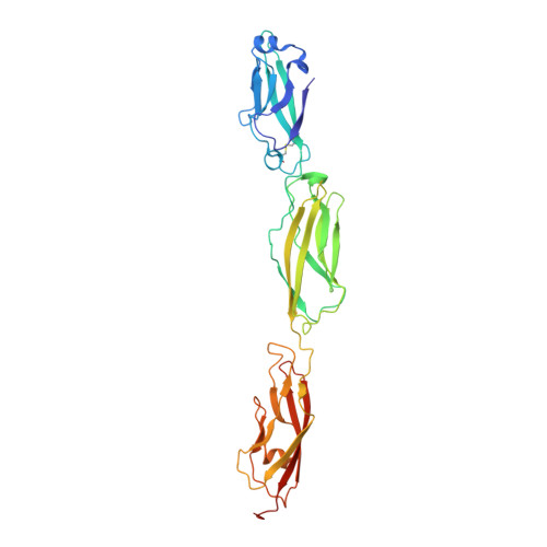
    Self-avoidance, a process preventing interactions of axons and dendrites from the same neuron during development, is mediated in vertebrates through the stochastic single-neuron expression of clustered protocadherin protein isoforms. Extracellular cadherin (EC) domains mediate isoform-specific homophilic binding between cells, conferring cell recognition through a poorly understood mechanism. Here, we report crystal structures for the EC1-EC3 domain regions from four protocadherin isoforms representing the α, β, and γ subfamilies. All are rod shaped and monomeric in solution. Biophysical measurements, cell aggregation assays, and computational docking reveal that trans binding between cells depends on the EC1-EC4 domains, which interact in an antiparallel orientation. We also show that the EC6 domains are required for the formation of cis-dimers. Overall, our results are consistent with a model in which protocadherin cis-dimers engage in a head-to-tail interaction between EC1-EC4 domains from apposed cell surfaces, possibly forming a zipper-like protein assembly, and thus providing a size-dependent self-recognition mechanism.


  • Organizational Affiliation

    Department of Biochemistry and Molecular Biophysics, Columbia University, New York, NY 10032, USA; Department of Systems Biology, Columbia University, New York, NY 10032, USA.


Macromolecules
Find similar proteins by:  (by identity cutoff)  |  3D Structure
Entity ID: 1
MoleculeChains Sequence LengthOrganismDetailsImage
Protein Pcdhb1322Mus musculusMutation(s): 0 
Gene Names: Pcdhb1
UniProt
Find proteins for Q91Y08 (Mus musculus)
Explore Q91Y08 
Go to UniProtKB:  Q91Y08
Entity Groups  
Sequence Clusters30% Identity50% Identity70% Identity90% Identity95% Identity100% Identity
UniProt GroupQ91Y08
Sequence Annotations
Expand
  • Reference Sequence
Oligosaccharides

Help

Entity ID: 2
MoleculeChains Length2D Diagram Glycosylation3D Interactions
beta-D-mannopyranose-(1-4)-2-acetamido-2-deoxy-beta-D-glucopyranose-(1-4)-[alpha-L-fucopyranose-(1-6)]2-acetamido-2-deoxy-beta-D-glucopyranose
B, C, D
4N-Glycosylation
Glycosylation Resources
GlyTouCan:  G32152BH
GlyCosmos:  G32152BH
GlyGen:  G32152BH
Experimental Data & Validation

Experimental Data

  • Method: X-RAY DIFFRACTION
  • Resolution: 3.30 Å
  • R-Value Free: 0.278 
  • R-Value Work: 0.231 
  • R-Value Observed: 0.234 
  • Space Group: I 21 21 21
Unit Cell:
Length ( Å )Angle ( ˚ )
a = 74.99α = 90
b = 106.52β = 90
c = 149.35γ = 90
Software Package:
Software NamePurpose
PHENIXrefinement
MOSFLMdata reduction
Aimlessdata scaling
PHASERphasing

Structure Validation

View Full Validation Report


Entry History 

Deposition Data

Revision History  (Full details and data files)

  • Version 1.0: 2015-10-28
    Type: Initial release
  • Version 1.1: 2015-11-04
    Changes: Database references
  • Version 2.0: 2020-07-29
    Type: Remediation
    Reason: Carbohydrate remediation
    Changes: Atomic model, Data collection, Derived calculations, Structure summary
  • Version 2.1: 2023-09-27
    Changes: Data collection, Database references, Derived calculations, Refinement description, Structure summary