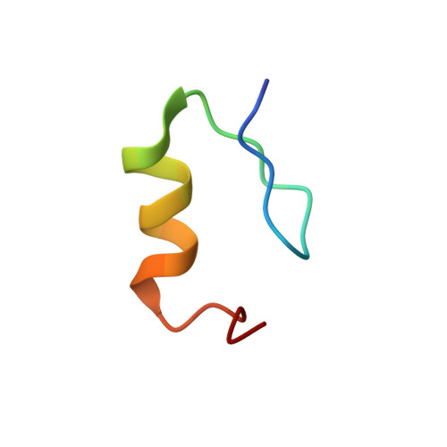
High-resolution three-dimensional structure of a single zinc finger from a human enhancer binding protein in solution.
Omichinski, J.G., Clore, G.M., Appella, E., Sakaguchi, K., Gronenborn, A.M.(1990) Biochemistry 29: 9324-9334
- PubMed: 2248949
- DOI: https://doi.org/10.1021/bi00492a004
- Primary Citation of Related Structures:
3ZNF, 4ZNF - PubMed Abstract:
The three-dimensional structure of a 30-residue synthetic peptide containing the carboxy-terminal "zinc finger" motif of a human enhancer binding protein has been determined by two-dimensional nuclear magnetic resonance (2D NMR) spectroscopy and hybrid distance geometry-dynamical simulated annealing calculations. The structure determination is based on 487 approximate interproton distance and 63 torsion angle (phi, psi, and chi 1) restraints. A total of 40 simulated annealing structures were calculated, and the atomic rms distribution about the mean coordinate positions (excluding residues 29 and 30 which are ill-defined) is 0.4 A for the backbone atoms, 0.8 A for all atoms, and 0.41 A for all atoms excluding the lysine and arginine side chains, which are disordered. The solution structure of the zinc finger consists of two irregular antiparallel beta-strands connected by an atypical turn (residues 3-12) and a classical alpha-helix (residues 14-24). The zinc is tetrahedrally coordinated to the sulfur atoms of two cysteines (Cys-5 and Cys-8) and to the N epsilon 2 atoms of two histidines (His-21 and His-27). The two cysteine residues are located in the turn connecting the two beta-strands (residues 5-8); one of the histidine ligands (His-21) is in the alpha-helix, while the second histidine (His-27) is at the end of a looplike structure (formed by the end of the alpha-helix and a turn). The general architecture is qualitatively similar to two previously determined low-resolution Cys2-His2 zinc finger structures, although distinct differences can be observed in the beta-strands and turn and in the region around the two histidines coordinated to zinc. Comparison of the overall polypeptide fold of the enhancer binding protein zinc finger with known structures in the crystallographic data base reveals a striking similarity to one region (residues 23-44) of the X-ray structure of proteinase inhibitor domain III of Japanese quail ovomucoid [Papamokos, E., Weber, E., Bode, W., Huber, R., Empie, M. W., Kato, I., & Laskowski, M. (1982) J. Mol. Biol. 158, 515-537], which could be superimposed with a backbone atomic rms difference of 0.95 A on residues 3-25 (excluding residue 6) of the zinc finger from the enhancer binding protein. The presence of structural homology between two proteins of very different function may indicate that the so-called zinc finger motif is not unique for a class of DNA binding proteins but may represent a general folding motif found in a variety of proteins irrespective of their function.
Organizational Affiliation:
Laboratory of Chemical Physics, National Institute of Diabetes and Digestive and Kidney Diseases, National Institutes of Health, Bethesda, Maryland 20892.