Sent packing: protein engineering generates a new crystal form of Pseudomonas aeruginosa DsbA1 with increased catalytic surface accessibility.
McMahon, R.M., Coincon, M., Tay, S., Heras, B., Morton, C.J., Scanlon, M.J., Martin, J.L.(2015) Acta Crystallogr D Biol Crystallogr 71: 2386-2395
- PubMed: 26627647
- DOI: https://doi.org/10.1107/S1399004715018519
- Primary Citation of Related Structures:
4ZL7, 4ZL8, 4ZL9 - PubMed Abstract:
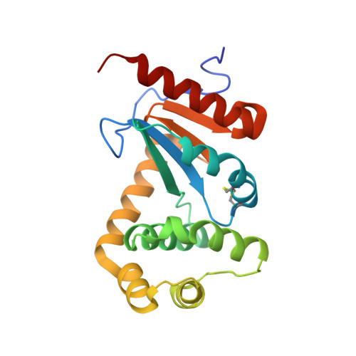
Pseudomonas aeruginosa is an opportunistic human pathogen for which new antimicrobial drug options are urgently sought. P. aeruginosa disulfide-bond protein A1 (PaDsbA1) plays a pivotal role in catalyzing the oxidative folding of multiple virulence proteins and as such holds great promise as a drug target. As part of a fragment-based lead discovery approach to PaDsbA1 inhibitor development, the identification of a crystal form of PaDsbA1 that was more suitable for fragment-soaking experiments was sought. A previously identified crystallization condition for this protein was unsuitable, as in this crystal form of PaDsbA1 the active-site surface loops are engaged in the crystal packing, occluding access to the target site. A single residue involved in crystal-packing interactions was substituted with an amino acid commonly found at this position in closely related enzymes, and this variant was successfully used to generate a new crystal form of PaDsbA1 in which the active-site surface is more accessible for soaking experiments. The PaDsbA1 variant displays identical redox character and in vitro activity to wild-type PaDsbA1 and is structurally highly similar. Two crystal structures of the PaDsbA1 variant were determined in complex with small molecules bound to the protein active site. These small molecules (MES, glycerol and ethylene glycol) were derived from the crystallization or cryoprotectant solutions and provide a proof of principle that the reported crystal form will be amenable to co-crystallization and soaking with small molecules designed to target the protein active-site surface.
Organizational Affiliation:
Institute for Molecular Bioscience, Division of Chemistry and Structural Biology, University of Queensland, 306 Carmody Road, Brisbane, Queensland 4072, Australia.