4ZI6

Crystal structure of leucine aminopeptidase from Helicobacter pylori


Experimental Data Snapshot

  • Method: X-RAY DIFFRACTION
  • Resolution: 2.00 Å
  • R-Value Free: 0.225 
  • R-Value Work: 0.171 
  • R-Value Observed: 0.173 

wwPDB Validation   3D Report Full Report


This is version 1.2 of the entry. See complete history


Literature

Structural basis for substrate specificity of Helicobacter pylori M17 aminopeptidase.

Modak, J.K.Rut, W.Wijeyewickrema, L.C.Pike, R.N.Drag, M.Roujeinikova, A.

(2016) Biochimie 121: 60-71

  • DOI: https://doi.org/10.1016/j.biochi.2015.11.021
  • Primary Citation of Related Structures:  
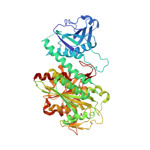
    4ZI6, 4ZLA

  • PubMed Abstract: 

    The M17 aminopeptidase from the carcinogenic gastric bacterium Helicobacter pylori (HpM17AP) is an important housekeeping enzyme involved in catabolism of endogenous and exogenous peptides. It is implicated in H. pylori defence against the human innate immune response and in the mechanism of metronidazole resistance. Bestatin inhibits HpM17AP and suppresses H. pylori growth. To address the structural basis of catalysis and inhibition of this enzyme, we have established its specificity towards the N-terminal amino acid of peptide substrates and determined the crystal structures of HpM17AP and its complex with bestatin. The position of the D-phenylalanine moiety of the inhibitor with respect to the active-site metal ions, bicarbonate ion and with respect to other M17 aminopeptidases suggested that this residue binds to the S1 subsite of HpM17AP. In contrast to most characterized M17 aminopeptidases, HpM17AP displays preference for L-Arg over L-Leu residues in peptide substrates. Compared to very similar homologues from other bacteria, a distinguishing feature of HpM17AP is a hydrophilic pocket at the end of the S1 subsite that is likely to accommodate the charged head group of the L-Arg residue of the substrate. The pocket is flanked by a sodium ion (not present in M17 aminopeptidases that show preference for L-Leu) and its coordinating water molecules. In addition, the structure suggests that variable loops at the entrance to, and in the middle of, the substrate-binding channel are important determinants of substrate specificity of M17 aminopeptidases.


  • Organizational Affiliation

    Infection and Immunity Program, Monash Biomedical Discovery Institute and Department of Microbiology, Monash University, Clayton, Victoria, Australia.


Macromolecules
Find similar proteins by:  (by identity cutoff)  |  3D Structure
Entity ID: 1
MoleculeChains Sequence LengthOrganismDetailsImage
Cytosol aminopeptidase
A, B, C, D, E
A, B, C, D, E, F
502Helicobacter pylori 26695Mutation(s): 0 
Gene Names: pepAHP_0570
EC: 3.4.11.1 (PDB Primary Data), 3.4.11.10 (PDB Primary Data)
UniProt
Find proteins for O25294 (Helicobacter pylori (strain ATCC 700392 / 26695))
Explore O25294 
Go to UniProtKB:  O25294
Entity Groups  
Sequence Clusters30% Identity50% Identity70% Identity90% Identity95% Identity100% Identity
UniProt GroupO25294
Sequence Annotations
Expand
  • Reference Sequence
Small Molecules
Ligands 3 Unique
IDChains Name / Formula / InChI Key2D Diagram3D Interactions
ZN
Query on ZN

Download Ideal Coordinates CCD File 
AA [auth F]
BA [auth F]
G [auth A]
H [auth A]
K [auth B]
AA [auth F],
BA [auth F],
G [auth A],
H [auth A],
K [auth B],
L [auth B],
O [auth C],
P [auth C],
S [auth D],
T [auth D],
W [auth E],
X [auth E]
ZINC ION
Zn
PTFCDOFLOPIGGS-UHFFFAOYSA-N
BCT
Query on BCT

Download Ideal Coordinates CCD File 
CA [auth F]
I [auth A]
M [auth B]
Q [auth C]
U [auth D]
CA [auth F],
I [auth A],
M [auth B],
Q [auth C],
U [auth D],
Y [auth E]
BICARBONATE ION
C H O3
BVKZGUZCCUSVTD-UHFFFAOYSA-M
NA
Query on NA

Download Ideal Coordinates CCD File 
DA [auth F]
J [auth A]
N [auth B]
R [auth C]
V [auth D]
DA [auth F],
J [auth A],
N [auth B],
R [auth C],
V [auth D],
Z [auth E]
SODIUM ION
Na
FKNQFGJONOIPTF-UHFFFAOYSA-N
Experimental Data & Validation

Experimental Data

  • Method: X-RAY DIFFRACTION
  • Resolution: 2.00 Å
  • R-Value Free: 0.225 
  • R-Value Work: 0.171 
  • R-Value Observed: 0.173 
  • Space Group: P 1
Unit Cell:
Length ( Å )Angle ( ˚ )
a = 96.31α = 75.26
b = 99.43β = 61.08
c = 99.68γ = 82.04
Software Package:
Software NamePurpose
SCALAdata scaling
PHENIXrefinement
PDB_EXTRACTdata extraction
MOSFLMdata reduction
PHASERphasing
Blu-Icedata collection

Structure Validation

View Full Validation Report


Entry History 

Deposition Data

Revision History  (Full details and data files)

  • Version 1.0: 2015-12-23
    Type: Initial release
  • Version 1.1: 2016-02-03
    Changes: Database references
  • Version 1.2: 2024-03-06
    Changes: Data collection, Database references, Derived calculations