4ZI4

YopH W354H Yersinia enterocolitica PTPase bond with Divanadate glycerol ester in the active site


Experimental Data Snapshot

  • Method: X-RAY DIFFRACTION
  • Resolution: 1.12 Å
  • R-Value Free: 0.159 
  • R-Value Work: 0.141 
  • R-Value Observed: 0.142 

wwPDB Validation   3D Report Full Report


Ligand Structure Quality Assessment 


This is version 1.5 of the entry. See complete history


Literature

Conservative Tryptophan Mutants of the Protein Tyrosine Phosphatase YopH Exhibit Impaired WPD-Loop Function and Crystallize with Divanadate Esters in Their Active Sites.

Moise, G.Gallup, N.M.Alexandrova, A.N.Hengge, A.C.Johnson, S.J.

(2015) Biochemistry 54: 6490-6500

  • DOI: https://doi.org/10.1021/acs.biochem.5b00496
  • Primary Citation of Related Structures:  
    4YAA, 4Z6B, 4ZI4, 4ZN5

  • PubMed Abstract: 

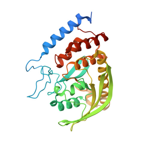
    Catalysis in protein tyrosine phosphatases (PTPs) involves movement of a protein loop called the WPD loop that brings a conserved aspartic acid into the active site to function as a general acid. Mutation of the tryptophan in the WPD loop of the PTP YopH to any other residue with a planar, aromatic side chain (phenylalanine, tyrosine, or histidine) disables general acid catalysis. Crystal structures reveal these conservative mutations leave this critical loop in a catalytically unproductive, quasi-open position. Although the loop positions in crystal structures are similar for all three conservative mutants, the reasons inhibiting normal loop closure differ for each mutant. In the W354F and W354Y mutants, steric clashes result from six-membered rings occupying the position of the five-membered ring of the native indole side chain. The histidine mutant dysfunction results from new hydrogen bonds stabilizing the unproductive position. The results demonstrate how even modest modifications can disrupt catalytically important protein dynamics. Crystallization of all the catalytically compromised mutants in the presence of vanadate gave rise to vanadate dimers at the active site. In W354Y and W354H, a divanadate ester with glycerol is observed. Such species have precedence in solution and are known from the small molecule crystal database. Such species have not been observed in the active site of a phosphatase, as a functional phosphatase would rapidly catalyze their decomposition. The compromised functionality of the mutants allows the trapping of species that undoubtedly form in solution and are capable of binding at the active sites of PTPs, and, presumably, other phosphatases. In addition to monomeric vanadate, such higher-order vanadium-based molecules are likely involved in the interaction of vanadate with PTPs in solution.


  • Organizational Affiliation

    Department of Chemistry and Biochemistry, Utah State University , Logan, Utah 84322-0300, United States.


Macromolecules
Find similar proteins by:  (by identity cutoff)  |  3D Structure
Entity ID: 1
MoleculeChains Sequence LengthOrganismDetailsImage
Tyrosine-protein phosphatase YopH306Yersinia enterocoliticaMutation(s): 2 
Gene Names: yopHyop51
EC: 3.1.3.48
UniProt
Find proteins for P15273 (Yersinia enterocolitica)
Explore P15273 
Go to UniProtKB:  P15273
Entity Groups  
Sequence Clusters30% Identity50% Identity70% Identity90% Identity95% Identity100% Identity
UniProt GroupP15273
Sequence Annotations
Expand
  • Reference Sequence
Small Molecules
Ligands 2 Unique
IDChains Name / Formula / InChI Key2D Diagram3D Interactions
DVG
Query on DVG

Download Ideal Coordinates CCD File 
B [auth A]Divanadate Glycerol ester
C3 H10 O8 V2
NROZHPLEEURAFR-ALWRQVICSA-J
VO4
Query on VO4

Download Ideal Coordinates CCD File 
C [auth A]VANADATE ION
O4 V
LSGOVYNHVSXFFJ-UHFFFAOYSA-N
Experimental Data & Validation

Experimental Data

  • Method: X-RAY DIFFRACTION
  • Resolution: 1.12 Å
  • R-Value Free: 0.159 
  • R-Value Work: 0.141 
  • R-Value Observed: 0.142 
  • Space Group: P 21 21 21
Unit Cell:
Length ( Å )Angle ( ˚ )
a = 52.888α = 90
b = 60.512β = 90
c = 89.358γ = 90
Software Package:
Software NamePurpose
PHENIXrefinement
HKL-2000data collection
SCALEPACKdata scaling
PDB_EXTRACTdata extraction
AUTOMARdata reduction
PHASERphasing

Structure Validation

View Full Validation Report


Ligand Structure Quality Assessment 


Entry History & Funding Information

Deposition Data


Funding OrganizationLocationGrant Number
National Institutes of Health/National Institute of General Medical Sciences (NIH/NIGMS)United StatesGM47297

Revision History  (Full details and data files)

  • Version 1.0: 2015-10-28
    Type: Initial release
  • Version 1.1: 2015-11-11
    Changes: Database references
  • Version 1.2: 2017-08-23
    Changes: Data collection, Database references, Derived calculations
  • Version 1.3: 2017-09-27
    Changes: Author supporting evidence
  • Version 1.4: 2019-12-25
    Changes: Author supporting evidence
  • Version 1.5: 2023-09-27
    Changes: Data collection, Database references, Refinement description