Structure and primase-mediated activation of a bacterial dodecameric replicative helicase.
Bazin, A., Cherrier, M.V., Gutsche, I., Timmins, J., Terradot, L.(2015) Nucleic Acids Res 43: 8564-8576
- PubMed: 26264665
- DOI: https://doi.org/10.1093/nar/gkv792
- Primary Citation of Related Structures:
4ZC0 - PubMed Abstract:
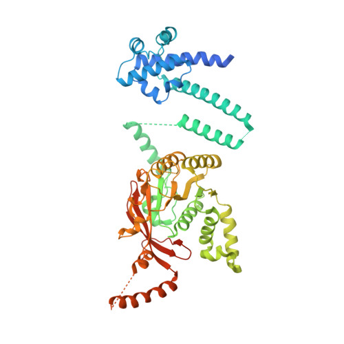
Replicative helicases are essential ATPases that unwind DNA to initiate chromosomal replication. While bacterial replicative DnaB helicases are hexameric, Helicobacter pylori DnaB (HpDnaB) was found to form double hexamers, similar to some archaeal and eukaryotic replicative helicases. Here we present a structural and functional analysis of HpDnaB protein during primosome formation. The crystal structure of the HpDnaB at 6.7 Å resolution reveals a dodecameric organization consisting of two hexamers assembled via their N-terminal rings in a stack-twisted mode. Using fluorescence anisotropy we show that HpDnaB dodecamer interacts with single-stranded DNA in the presence of ATP but has a low DNA unwinding activity. Multi-angle light scattering and small angle X-ray scattering demonstrate that interaction with the DnaG primase helicase-binding domain dissociates the helicase dodecamer into single ringed primosomes. Functional assays on the proteins and associated complexes indicate that these single ringed primosomes are the most active form of the helicase for ATP hydrolysis, DNA binding and unwinding. These findings shed light onto an activation mechanism of HpDnaB by the primase that might be relevant in other bacteria and possibly other organisms exploiting dodecameric helicases for DNA replication.
Organizational Affiliation:
CNRS, UMR 5086 Bases Moléculaires et Structurales de Systèmes Infectieux, Institut de Biologie et Chimie des Protéines, 7 Passage du Vercors, F-69367, Lyon, France. Université de Lyon, F-69622, Lyon, France; Université Claude Bernard Lyon 1, F-69622, Villeurbanne, France.