4ZA3

Structural studies on a non-toxic homologue of type II RIPs from Momordica charantia (bitter gourd)-Native-3


Experimental Data Snapshot

  • Method: X-RAY DIFFRACTION
  • Resolution: 1.67 Å
  • R-Value Free: 0.183 
  • R-Value Work: 0.160 
  • R-Value Observed: 0.161 

wwPDB Validation   3D Report Full Report


Ligand Structure Quality Assessment 


This is version 2.1 of the entry. See complete history


Literature

Structural studies on a non-toxic homologue of type II RIPs from bitter gourd: Molecular basis of non-toxicity, conformational selection and glycan structure.

Chandran, T.Sharma, A.Vijayan, M.

(2015) J Biosci 40: 929-941

  • DOI: https://doi.org/10.1007/s12038-015-9573-x
  • Primary Citation of Related Structures:  
    4Z8S, 4Z9W, 4ZA3, 4ZBV, 4ZFU, 4ZFW, 4ZFY, 4ZGR, 4ZLB

  • PubMed Abstract: 

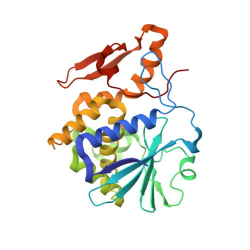
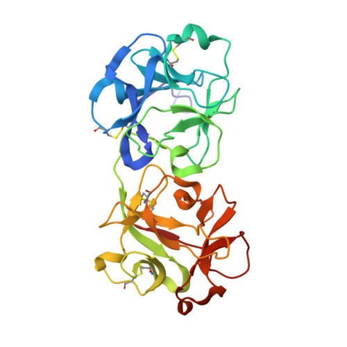
    The structures of nine independent crystals of bitter gourd seed lectin (BGSL), a non-toxic homologue of type II RIPs, and its sugar complexes have been determined. The four-chain, two-fold symmetric, protein is made up of two identical two-chain modules, each consisting of a catalytic chain and a lectin chain, connected by a disulphide bridge. The lectin chain is made up of two domains. Each domain carries a carbohydrate binding site in type II RIPs of known structure. BGSL has a sugar binding site only on one domain, thus impairing its interaction at the cell surface. The adenine binding site in the catalytic chain is defective. Thus, defects in sugar binding as well as adenine binding appear to contribute to the non-toxicity of the lectin. The plasticity of the molecule is mainly caused by the presence of two possible well defined conformations of a surface loop in the lectin chain. One of them is chosen in the sugar complexes, in a case of conformational selection, as the chosen conformation facilitates an additional interaction with the sugar, involving an arginyl residue in the loop. The N-glycosylation of the lectin involves a plant-specific glycan while that in toxic type II RIPs of known structure involves a glycan which is animal as well as plant specific.


  • Organizational Affiliation

    Molecular Biophysics Unit, Indian Institute of Science, Bangalore 560012, India.


Macromolecules
Find similar proteins by:  (by identity cutoff)  |  3D Structure
Entity ID: 1
MoleculeChains Sequence LengthOrganismDetailsImage
rRNA N-glycosidase247Momordica charantiaMutation(s): 0 
EC: 3.2.2.22
UniProt
Find proteins for B7X8M2 (Momordica charantia)
Explore B7X8M2 
Go to UniProtKB:  B7X8M2
Entity Groups  
Sequence Clusters30% Identity50% Identity70% Identity90% Identity95% Identity100% Identity
UniProt GroupB7X8M2
Sequence Annotations
Expand
  • Reference Sequence
Find similar proteins by:  (by identity cutoff)  |  3D Structure
Entity ID: 2
MoleculeChains Sequence LengthOrganismDetailsImage
rRNA N-glycosidase260Momordica charantiaMutation(s): 0 
EC: 3.2.2.22
UniProt
Find proteins for B7X8M2 (Momordica charantia)
Explore B7X8M2 
Go to UniProtKB:  B7X8M2
Entity Groups  
Sequence Clusters30% Identity50% Identity70% Identity90% Identity95% Identity100% Identity
UniProt GroupB7X8M2
Sequence Annotations
Expand
  • Reference Sequence
Oligosaccharides

Help

Entity ID: 3
MoleculeChains Length2D Diagram Glycosylation3D Interactions
alpha-L-fucopyranose-(1-3)-2-acetamido-2-deoxy-beta-D-glucopyranose
C
2N-Glycosylation
Glycosylation Resources
GlyTouCan:  G80587NA
GlyCosmos:  G80587NA
GlyGen:  G80587NA
Entity ID: 4
MoleculeChains Length2D Diagram Glycosylation3D Interactions
beta-D-mannopyranose-(1-4)-2-acetamido-2-deoxy-beta-D-glucopyranose
D
2N/A
Glycosylation Resources
GlyTouCan:  G90333CG
GlyCosmos:  G90333CG
GlyGen:  G90333CG
Entity ID: 5
MoleculeChains Length2D Diagram Glycosylation3D Interactions
2-acetamido-2-deoxy-beta-D-glucopyranose-(1-4)-2-acetamido-2-deoxy-beta-D-glucopyranose
E
2N-Glycosylation
Glycosylation Resources
GlyTouCan:  G42666HT
GlyCosmos:  G42666HT
GlyGen:  G42666HT
Small Molecules
Ligands 3 Unique
IDChains Name / Formula / InChI Key2D Diagram3D Interactions
NAG
Query on NAG

Download Ideal Coordinates CCD File 
H [auth A]2-acetamido-2-deoxy-beta-D-glucopyranose
C8 H15 N O6
OVRNDRQMDRJTHS-FMDGEEDCSA-N
GOL
Query on GOL

Download Ideal Coordinates CCD File 
M [auth B]GLYCEROL
C3 H8 O3
PEDCQBHIVMGVHV-UHFFFAOYSA-N
EDO
Query on EDO

Download Ideal Coordinates CCD File 
F [auth A]
G [auth A]
I [auth B]
J [auth B]
K [auth B]
F [auth A],
G [auth A],
I [auth B],
J [auth B],
K [auth B],
L [auth B]
1,2-ETHANEDIOL
C2 H6 O2
LYCAIKOWRPUZTN-UHFFFAOYSA-N
Experimental Data & Validation

Experimental Data

  • Method: X-RAY DIFFRACTION
  • Resolution: 1.67 Å
  • R-Value Free: 0.183 
  • R-Value Work: 0.160 
  • R-Value Observed: 0.161 
  • Space Group: P 21 21 2
Unit Cell:
Length ( Å )Angle ( ˚ )
a = 141.4α = 90
b = 119.59β = 90
c = 44.86γ = 90
Software Package:
Software NamePurpose
REFMACrefinement
MOSFLMdata collection
SCALAdata scaling
PDB_EXTRACTdata extraction

Structure Validation

View Full Validation Report


Ligand Structure Quality Assessment 


Entry History & Funding Information

Deposition Data


Funding OrganizationLocationGrant Number
Science and Engineering Research Board, Department of Science and TechnologyIndia--

Revision History  (Full details and data files)

  • Version 1.0: 2016-03-23
    Type: Initial release
  • Version 1.1: 2017-09-27
    Changes: Data collection, Database references, Derived calculations
  • Version 1.2: 2019-10-09
    Changes: Data collection, Database references
  • Version 2.0: 2020-07-29
    Type: Remediation
    Reason: Carbohydrate remediation
    Changes: Advisory, Atomic model, Data collection, Derived calculations, Structure summary
  • Version 2.1: 2023-11-08
    Changes: Data collection, Database references, Derived calculations, Refinement description, Structure summary