4Z7H

Crystal structure of human IRE1 cytoplasmic kinase-RNase region - complex with imidazopyridine compound 3


Experimental Data Snapshot

  • Method: X-RAY DIFFRACTION
  • Resolution: 2.90 Å
  • R-Value Free: 0.225 
  • R-Value Work: 0.200 
  • R-Value Observed: 0.202 

wwPDB Validation   3D Report Full Report


Ligand Structure Quality Assessment 


This is version 1.3 of the entry. See complete history


Literature

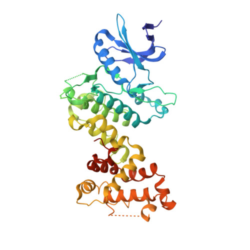
Molecular mechanisms of human IRE1 activation through dimerization and ligand binding.

Joshi, A.Newbatt, Y.McAndrew, P.C.Stubbs, M.Burke, R.Richards, M.W.Bhatia, C.Caldwell, J.J.McHardy, T.Collins, I.Bayliss, R.

(2015) Oncotarget 6: 13019-13035

  • DOI: https://doi.org/10.18632/oncotarget.3864
  • Primary Citation of Related Structures:  
    4Z7G, 4Z7H

  • PubMed Abstract: 

    IRE1 transduces the unfolded protein response by splicing XBP1 through its C-terminal cytoplasmic kinase-RNase region. IRE1 autophosphorylation is coupled to RNase activity through formation of a back-to-back dimer, although the conservation of the underlying molecular mechanism is not clear from existing structures. We have crystallized human IRE1 in a back-to-back conformation only previously seen for the yeast homologue. In our structure the kinase domain appears primed for catalysis but the RNase domains are disengaged. Structure-function analysis reveals that IRE1 is autoinhibited through a Tyr-down mechanism related to that found in the unrelated Ser/Thr protein kinase Nek7. We have developed a compound that potently inhibits human IRE1 kinase activity while stimulating XBP1 splicing. A crystal structure of the inhibitor bound to IRE1 shows an increased ordering of the kinase activation loop. The structures of hIRE in apo and ligand-bound forms are consistent with a previously proposed model of IRE1 regulation in which formation of a back-to-back dimer coupled to adoption of a kinase-active conformation drive RNase activation. The structures provide opportunities for structure-guided design of IRE1 inhibitors.


  • Organizational Affiliation

    Department of Biochemistry, University of Leicester, Leicester, United Kingdom.


Macromolecules
Find similar proteins by:  (by identity cutoff)  |  3D Structure
Entity ID: 1
MoleculeChains Sequence LengthOrganismDetailsImage
Serine/threonine-protein kinase/endoribonuclease IRE1
A, B
416Homo sapiensMutation(s): 0 
Gene Names: ERN1IRE1
EC: 2.7.11.1 (PDB Primary Data), 3.1.26 (PDB Primary Data)
UniProt & NIH Common Fund Data Resources
Find proteins for O75460 (Homo sapiens)
Explore O75460 
Go to UniProtKB:  O75460
PHAROS:  O75460
GTEx:  ENSG00000178607 
Entity Groups  
Sequence Clusters30% Identity50% Identity70% Identity90% Identity95% Identity100% Identity
UniProt GroupO75460
Sequence Annotations
Expand
  • Reference Sequence
Small Molecules
Experimental Data & Validation

Experimental Data

  • Method: X-RAY DIFFRACTION
  • Resolution: 2.90 Å
  • R-Value Free: 0.225 
  • R-Value Work: 0.200 
  • R-Value Observed: 0.202 
  • Space Group: P 21 21 21
Unit Cell:
Length ( Å )Angle ( ˚ )
a = 78.678α = 90
b = 81.957β = 90
c = 168.682γ = 90
Software Package:
Software NamePurpose
BUSTERrefinement
XDSdata reduction
Aimlessdata scaling

Structure Validation

View Full Validation Report


Ligand Structure Quality Assessment 


Entry History & Funding Information

Deposition Data


Funding OrganizationLocationGrant Number
Cancer Research UKUnited KingdomC24461/A13231

Revision History  (Full details and data files)

  • Version 1.0: 2015-05-27
    Type: Initial release
  • Version 1.1: 2015-07-01
    Changes: Database references
  • Version 1.2: 2019-10-09
    Changes: Data collection, Database references
  • Version 1.3: 2024-01-10
    Changes: Data collection, Database references, Refinement description