


Crystal structure of urease from Yersinia enterocolitica
Studer, G., Jakob, R.P., Mahi, M.A., Wiesand, U., Schwede, T., Maier, T.To be published.
Experimental Data Snapshot
wwPDB Validation 3D Report Full Report

Entity ID: 1 | |||||
|---|---|---|---|---|---|
| Molecule | Chains | Sequence Length | Organism | Details | Image |
| Urease subunit gamma | 100 | Yersinia enterocolitica W22703 | Mutation(s): 0 EC: 3.5.1.5 | 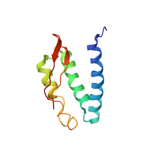 | |
UniProt | |||||
Find proteins for F4MWM9 (Yersinia enterocolitica W22703) Explore F4MWM9 Go to UniProtKB: F4MWM9 | |||||
Entity Groups | |||||
| Sequence Clusters | 30% Identity50% Identity70% Identity90% Identity95% Identity100% Identity | ||||
| UniProt Group | F4MWM9 | ||||
Sequence AnnotationsExpand | |||||
| |||||
Entity ID: 2 | |||||
|---|---|---|---|---|---|
| Molecule | Chains | Sequence Length | Organism | Details | Image |
| Urease subunit beta | 164 | Yersinia enterocolitica W22703 | Mutation(s): 0 EC: 3.5.1.5 | 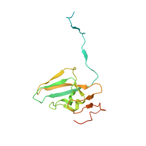 | |
UniProt | |||||
Find proteins for F4MWM8 (Yersinia enterocolitica W22703) Explore F4MWM8 Go to UniProtKB: F4MWM8 | |||||
Entity Groups | |||||
| Sequence Clusters | 30% Identity50% Identity70% Identity90% Identity95% Identity100% Identity | ||||
| UniProt Group | F4MWM8 | ||||
Sequence AnnotationsExpand | |||||
| |||||
Entity ID: 3 | |||||
|---|---|---|---|---|---|
| Molecule | Chains | Sequence Length | Organism | Details | Image |
| Urease subunit alpha | 572 | Yersinia enterocolitica W22703 | Mutation(s): 0 EC: 3.5.1.5 | 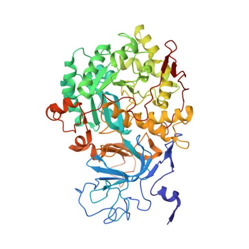 | |
UniProt | |||||
Find proteins for F4MWM7 (Yersinia enterocolitica W22703) Explore F4MWM7 Go to UniProtKB: F4MWM7 | |||||
Entity Groups | |||||
| Sequence Clusters | 30% Identity50% Identity70% Identity90% Identity95% Identity100% Identity | ||||
| UniProt Group | F4MWM7 | ||||
Sequence AnnotationsExpand | |||||
| |||||
| Ligands 1 Unique | |||||
|---|---|---|---|---|---|
| ID | Chains | Name / Formula / InChI Key | 2D Diagram | 3D Interactions | |
| NI Query on NI | M [auth C] N [auth C] O [auth F] P [auth F] Q [auth I] | NICKEL (II) ION Ni VEQPNABPJHWNSG-UHFFFAOYSA-N |  | ||
| Length ( Å ) | Angle ( ˚ ) |
|---|---|
| a = 157.2 | α = 90 |
| b = 157.2 | β = 90 |
| c = 774.62 | γ = 120 |
| Software Name | Purpose |
|---|---|
| BUSTER | refinement |
| XDS | data reduction |
| SCALA | data scaling |
| PHASER | phasing |
RCSB PDB (citation) is hosted by
RCSB PDB is a member of the

