Biochemical and Structural Characterization of Enolase from Chloroflexus aurantiacus: Evidence for a Thermophilic Origin.
Zadvornyy, O.A., Boyd, E.S., Posewitz, M.C., Zorin, N.A., Peters, J.W.(2015) Front Bioeng Biotechnol 3: 74-74
- PubMed: 26082925
- DOI: https://doi.org/10.3389/fbioe.2015.00074
- Primary Citation of Related Structures:
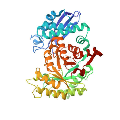
4YWS, 4Z17, 4Z1Y - PubMed Abstract:
Enolase catalyzes the conversion of 2-phosphoglycerate to phosphoenolpyruvate during both glycolysis and gluconeogenesis, and is required by all three domains of life. Here, we report the purification and biochemical and structural characterization of enolase from Chloroflexus aurantiacus, a thermophilic anoxygenic phototroph affiliated with the green non-sulfur bacteria. The protein was purified as a homodimer with a subunit molecular weight of 46 kDa. The temperature optimum for enolase catalysis was 80°C, close to the measured thermal stability of the protein which was determined to be 75°C, while the pH optimum for enzyme activity was 6.5. The specific activities of purified enolase determined at 25 and 80°C were 147 and 300 U mg(-1) of protein, respectively. K m values for the 2-phosphoglycerate/phosphoenolpyruvate reaction determined at 25 and 80°C were 0.16 and 0.03 mM, respectively. The K m values for Mg(2+) binding at these temperatures were 2.5 and 1.9 mM, respectively. When compared to enolase from mesophiles, the biochemical and structural properties of enolase from C. aurantiacus are consistent with this being thermally adapted. These data are consistent with the results of our phylogenetic analysis of enolase, which reveal that enolase has a thermophilic origin.
Organizational Affiliation:
Department of Chemistry and Biochemistry, Montana State University , Bozeman, MT , USA ; Institute of Basic Biological Problems, Russian Academy of Sciences , Pushchino , Russia.