4YV9

X-ray crystal structure of Streptococcus dysgalactiae SHP pheromone receptor Rgg2


Experimental Data Snapshot

  • Method: X-RAY DIFFRACTION
  • Resolution: 1.95 Å
  • R-Value Free: 0.213 
  • R-Value Work: 0.188 
  • R-Value Observed: 0.189 

wwPDB Validation   3D Report Full Report


This is version 1.3 of the entry. See complete history


Literature

Rgg protein structure-function and inhibition by cyclic peptide compounds.

Parashar, V.Aggarwal, C.Federle, M.J.Neiditch, M.B.

(2015) Proc Natl Acad Sci U S A 112: 5177-5182

  • DOI: https://doi.org/10.1073/pnas.1500357112
  • Primary Citation of Related Structures:  
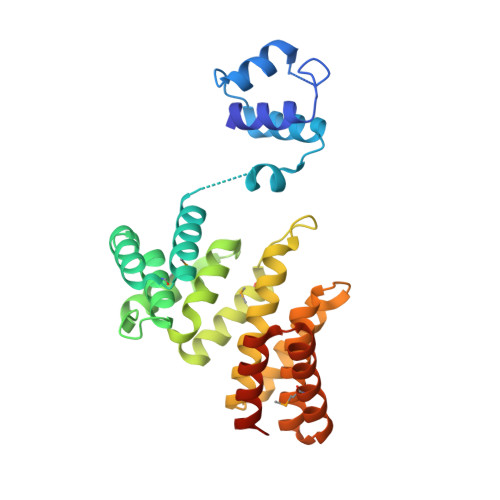
    4YV6, 4YV9

  • PubMed Abstract: 

    Peptide pheromone cell-cell signaling (quorum sensing) regulates the expression of diverse developmental phenotypes (including virulence) in Firmicutes, which includes common human pathogens, e.g., Streptococcus pyogenes and Streptococcus pneumoniae. Cytoplasmic transcription factors known as "Rgg proteins" are peptide pheromone receptors ubiquitous in Firmicutes. Here we present X-ray crystal structures of a Streptococcus Rgg protein alone and in complex with a tight-binding signaling antagonist, the cyclic undecapeptide cyclosporin A. To our knowledge, these represent the first Rgg protein X-ray crystal structures. Based on the results of extensive structure-function analysis, we reveal the peptide pheromone-binding site and the mechanism by which cyclosporin A inhibits activation of the peptide pheromone receptor. Guided by the Rgg-cyclosporin A complex structure, we predicted that the nonimmunosuppressive cyclosporin A analog valspodar would inhibit Rgg activation. Indeed, we found that, like cyclosporin A, valspodar inhibits peptide pheromone activation of conserved Rgg proteins in medically relevant Streptococcus species. Finally, the crystal structures presented here revealed that the Rgg protein DNA-binding domains are covalently linked across their dimerization interface by a disulfide bond formed by a highly conserved cysteine. The DNA-binding domain dimerization interface observed in our structures is essentially identical to the interfaces previously described for other members of the XRE DNA-binding domain family, but the presence of an intermolecular disulfide bond buried in this interface appears to be unique. We hypothesize that this disulfide bond may, under the right conditions, affect Rgg monomer-dimer equilibrium, stabilize Rgg conformation, or serve as a redox-sensitive switch.


  • Organizational Affiliation

    Department of Microbiology, Biochemistry, and Molecular Genetics, New Jersey Medical School, Rutgers, The State University of New Jersey, Newark, NJ 07103; and.


Macromolecules
Find similar proteins by:  (by identity cutoff)  |  3D Structure
Entity ID: 1
MoleculeChains Sequence LengthOrganismDetailsImage
Transcriptional regulator
A, B, C, D
284Streptococcus dysgalactiaeMutation(s): 0 
Gene Names: mutR
UniProt
Find proteins for A0A0J9X288 (Streptococcus dysgalactiae)
Explore A0A0J9X288 
Go to UniProtKB:  A0A0J9X288
Entity Groups  
Sequence Clusters30% Identity50% Identity70% Identity90% Identity95% Identity100% Identity
UniProt GroupA0A0J9X288
Sequence Annotations
Expand
  • Reference Sequence

Find similar proteins by:  Sequence   |   3D Structure  

Entity ID: 2
MoleculeChains Sequence LengthOrganismDetailsImage
Cyclosporin A
E, F, G, H
11Trichoderma polysporumMutation(s): 0 
Entity Groups  
Sequence Clusters30% Identity50% Identity70% Identity90% Identity95% Identity100% Identity
Sequence Annotations
Expand
  • Reference Sequence
Small Molecules
Ligands 1 Unique
IDChains Name / Formula / InChI Key2D Diagram3D Interactions
SO4
Query on SO4

Download Ideal Coordinates CCD File 
I [auth A]
J [auth A]
K [auth A]
L [auth A]
M [auth B]
I [auth A],
J [auth A],
K [auth A],
L [auth A],
M [auth B],
N [auth B],
O [auth C],
P [auth C],
Q [auth C],
R [auth D],
S [auth D],
T [auth D],
U [auth D]
SULFATE ION
O4 S
QAOWNCQODCNURD-UHFFFAOYSA-L
Modified Residues  6 Unique
IDChains TypeFormula2D DiagramParent
MSE
Query on MSE
A, B, C, D
L-PEPTIDE LINKINGC5 H11 N O2 SeMET
ABA
Query on ABA
E, F, G, H
L-PEPTIDE LINKINGC4 H9 N O2ALA
BMT
Query on BMT
E, F, G, H
L-PEPTIDE LINKINGC10 H19 N O3THR
MLE
Query on MLE
E, F, G, H
L-PEPTIDE LINKINGC7 H15 N O2LEU
MVA
Query on MVA
E, F, G, H
L-PEPTIDE LINKINGC6 H13 N O2VAL
SAR
Query on SAR
E, F, G, H
PEPTIDE LINKINGC3 H7 N O2GLY
Experimental Data & Validation

Experimental Data

  • Method: X-RAY DIFFRACTION
  • Resolution: 1.95 Å
  • R-Value Free: 0.213 
  • R-Value Work: 0.188 
  • R-Value Observed: 0.189 
  • Space Group: P 1 21 1
Unit Cell:
Length ( Å )Angle ( ˚ )
a = 68.388α = 90
b = 99.427β = 97
c = 99.882γ = 90
Software Package:
Software NamePurpose
PHENIXrefinement
HKL-2000data reduction
HKL-2000data scaling
PHENIXphasing

Structure Validation

View Full Validation Report


Entry History 

Deposition Data

Revision History  (Full details and data files)

  • Version 1.0: 2015-04-08
    Type: Initial release
  • Version 1.1: 2015-04-22
    Changes: Database references
  • Version 1.2: 2015-05-06
    Changes: Database references
  • Version 1.3: 2017-11-01
    Changes: Author supporting evidence, Database references, Derived calculations, Source and taxonomy