4YS1

Human Aldose Reductase complexed with a ligand with an IDD structure (2) at 1.07 A.


Experimental Data Snapshot

  • Method: X-RAY DIFFRACTION
  • Resolution: 1.07 Å
  • R-Value Free: 0.148 
  • R-Value Work: 0.132 
  • R-Value Observed: 0.132 

wwPDB Validation   3D Report Full Report


Ligand Structure Quality Assessment 


This is version 1.2 of the entry. See complete history


Literature

Price for Opening the Transient Specificity Pocket in Human Aldose Reductase upon Ligand Binding: Structural, Thermodynamic, Kinetic, and Computational Analysis.

Rechlin, C.Scheer, F.Terwesten, F.Wulsdorf, T.Pol, E.Fridh, V.Toth, P.Diederich, W.E.Heine, A.Klebe, G.

(2017) ACS Chem Biol 12: 1397-1415

  • DOI: https://doi.org/10.1021/acschembio.7b00062
  • Primary Citation of Related Structures:  
    4YS1

  • PubMed Abstract: 

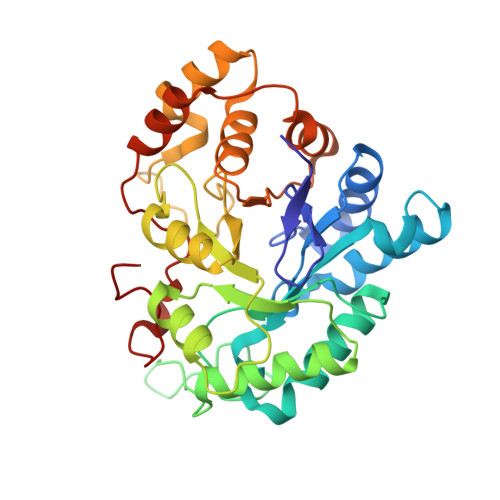
    Insights into the thermodynamic and kinetic signature of the transient opening of a protein-binding pocket resulting from accommodation of suitable substituents attached to a given parent ligand scaffold are presented. As a target, we selected human aldose reductase, an enzyme involved in the development of late-stage diabetic complications. To recognize a large scope of substrate molecules, this reductase opens a transient specificity pocket. The pocket-opening step was studied by X-ray crystallography, microcalorimetry, and surface plasmon resonance using a narrow series of 2-carbamoyl-phenoxy-acetic acid derivatives. Molecular dynamics simulations suggest that pocket opening occurs only once an appropriate substituent is attached to the parent scaffold. Transient pocket opening of the uncomplexed protein is hardly recorded. Hydration-site analysis suggests that up to five water molecules entering the opened pocket cannot stabilize this state. Sole substitution with a benzyl group stabilizes the opened state, and the energetic barrier for opening is estimated to be ∼5 kJ/mol. Additional decoration of the pocket-opening benzyl substituent with a nitro group results in a huge enthalpy-driven potency increase; on the other hand, an isosteric carboxylic acid group reduces the potency 1000-fold, and binding occurs without pocket opening. We suggest a ligand induced-fit mechanism for the pocket-opening step, which, however, does not represent the rate-determining step in binding kinetics.


  • Organizational Affiliation

    Institut für Pharmazeutische Chemie, Philipps-Universität Marburg , Marbacher Weg 6, D-35032 Marburg, Germany.


Macromolecules
Find similar proteins by:  (by identity cutoff)  |  3D Structure
Entity ID: 1
MoleculeChains Sequence LengthOrganismDetailsImage
Aldose reductase316Homo sapiensMutation(s): 0 
Gene Names: AKR1B1ALDR1
EC: 1.1.1.21
UniProt & NIH Common Fund Data Resources
Find proteins for P15121 (Homo sapiens)
Explore P15121 
Go to UniProtKB:  P15121
PHAROS:  P15121
GTEx:  ENSG00000085662 
Entity Groups  
Sequence Clusters30% Identity50% Identity70% Identity90% Identity95% Identity100% Identity
UniProt GroupP15121
Sequence Annotations
Expand
  • Reference Sequence
Small Molecules
Ligands 2 Unique
IDChains Name / Formula / InChI Key2D Diagram3D Interactions
NAP
Query on NAP

Download Ideal Coordinates CCD File 
B [auth A]NADP NICOTINAMIDE-ADENINE-DINUCLEOTIDE PHOSPHATE
C21 H28 N7 O17 P3
XJLXINKUBYWONI-NNYOXOHSSA-N
4G7
Query on 4G7

Download Ideal Coordinates CCD File 
C [auth A]3-({[2-(carboxymethoxy)-4-fluorobenzoyl]amino}methyl)benzoic acid
C17 H14 F N O6
NEABINVPHSWADT-UHFFFAOYSA-N
Binding Affinity Annotations 
IDSourceBinding Affinity
4G7 BindingDB:  4YS1 -TΔS: -1.10e+1 (kJ/mol) from 1 assay(s)
ΔH: -3.64e+1 (kJ/mol) from 1 assay(s)
ΔG: -2.54e+1 (kJ/mol) from 1 assay(s)
Experimental Data & Validation

Experimental Data

  • Method: X-RAY DIFFRACTION
  • Resolution: 1.07 Å
  • R-Value Free: 0.148 
  • R-Value Work: 0.132 
  • R-Value Observed: 0.132 
  • Space Group: P 1 21 1
Unit Cell:
Length ( Å )Angle ( ˚ )
a = 49.312α = 90
b = 66.62β = 92.15
c = 47.309γ = 90
Software Package:
Software NamePurpose
PHENIXrefinement
XDSdata reduction
PHASERphasing
XDSdata scaling

Structure Validation

View Full Validation Report


Ligand Structure Quality Assessment 


Entry History & Funding Information

Deposition Data


Funding OrganizationLocationGrant Number
European Union268145-DrugProfilBind

Revision History  (Full details and data files)

  • Version 1.0: 2016-03-23
    Type: Initial release
  • Version 1.1: 2017-12-06
    Changes: Database references
  • Version 1.2: 2024-01-10
    Changes: Data collection, Database references, Refinement description