4YMK

Crystal Structure of Stearoyl-Coenzyme A Desaturase 1


Experimental Data Snapshot

  • Method: X-RAY DIFFRACTION
  • Resolution: 2.60 Å
  • R-Value Free: 0.235 
  • R-Value Work: 0.203 
  • R-Value Observed: 0.204 

wwPDB Validation   3D Report Full Report


Ligand Structure Quality Assessment 


This is version 1.7 of the entry. See complete history


Literature

X-ray structure of a mammalian stearoyl-CoA desaturase.

Bai, Y.McCoy, J.G.Levin, E.J.Sobrado, P.Rajashankar, K.R.Fox, B.G.Zhou, M.

(2015) Nature 524: 252-256

  • DOI: https://doi.org/10.1038/nature14549
  • Primary Citation of Related Structures:  
    4YMK

  • PubMed Abstract: 

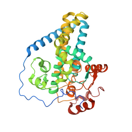
    Stearoyl-CoA desaturase (SCD) is conserved in all eukaryotes and introduces the first double bond into saturated fatty acyl-CoAs. Because the monounsaturated products of SCD are key precursors of membrane phospholipids, cholesterol esters and triglycerides, SCD is pivotal in fatty acid metabolism. Humans have two SCD homologues (SCD1 and SCD5), while mice have four (SCD1-SCD4). SCD1-deficient mice do not become obese or diabetic when fed a high-fat diet because of improved lipid metabolic profiles and insulin sensitivity. Thus, SCD1 is a pharmacological target in the treatment of obesity, diabetes and other metabolic diseases. SCD1 is an integral membrane protein located in the endoplasmic reticulum, and catalyses the formation of a cis-double bond between the ninth and tenth carbons of stearoyl- or palmitoyl-CoA. The reaction requires molecular oxygen, which is activated by a di-iron centre, and cytochrome b5, which regenerates the di-iron centre. To understand better the structural basis of these characteristics of SCD function, here we crystallize and solve the structure of mouse SCD1 bound to stearoyl-CoA at 2.6 Å resolution. The structure shows a novel fold comprising four transmembrane helices capped by a cytosolic domain, and a plausible pathway for lateral substrate access and product egress. The acyl chain of the bound stearoyl-CoA is enclosed in a tunnel buried in the cytosolic domain, and the geometry of the tunnel and the conformation of the bound acyl chain provide a structural basis for the regioselectivity and stereospecificity of the desaturation reaction. The dimetal centre is coordinated by a unique spacial arrangement of nine conserved histidine residues that implies a potentially novel mechanism for oxygen activation. The structure also illustrates a possible route for electron transfer from cytochrome b5 to the di-iron centre.


  • Organizational Affiliation

    Verna and Marrs McLean Department of Biochemistry and Molecular Biology, Baylor College of Medicine, Houston, Texas 77030, USA.


Macromolecules
Find similar proteins by:  (by identity cutoff)  |  3D Structure
Entity ID: 1
MoleculeChains Sequence LengthOrganismDetailsImage
Acyl-CoA desaturase 1A,
B [auth D]
339Mus musculusMutation(s): 0 
Gene Names: Scd1
EC: 1.14.19.1
Membrane Entity: Yes 
UniProt
Find proteins for P13516 (Mus musculus)
Explore P13516 
Go to UniProtKB:  P13516
Entity Groups  
Sequence Clusters30% Identity50% Identity70% Identity90% Identity95% Identity100% Identity
UniProt GroupP13516
Sequence Annotations
Expand
  • Reference Sequence
Small Molecules
Ligands 3 Unique
IDChains Name / Formula / InChI Key2D Diagram3D Interactions
ST9
Query on ST9

Download Ideal Coordinates CCD File 
E [auth A],
I [auth D]
STEAROYL-COENZYME A
C39 H70 N7 O17 P3 S
SIARJEKBADXQJG-LFZQUHGESA-N
MPG
Query on MPG

Download Ideal Coordinates CCD File 
F [auth A],
J [auth D]
[(Z)-octadec-9-enyl] (2R)-2,3-bis(oxidanyl)propanoate
C21 H40 O4
JPJYKWFFJCWMPK-GDCKJWNLSA-N
ZN
Query on ZN

Download Ideal Coordinates CCD File 
C [auth A],
D [auth A],
G [auth D],
H [auth D]
ZINC ION
Zn
PTFCDOFLOPIGGS-UHFFFAOYSA-N
Experimental Data & Validation

Experimental Data

  • Method: X-RAY DIFFRACTION
  • Resolution: 2.60 Å
  • R-Value Free: 0.235 
  • R-Value Work: 0.203 
  • R-Value Observed: 0.204 
  • Space Group: P 21 21 21
Unit Cell:
Length ( Å )Angle ( ˚ )
a = 77.061α = 90
b = 113.766β = 90
c = 141.698γ = 90
Software Package:
Software NamePurpose
PHENIXrefinement
XDSdata scaling
Aimlessdata scaling
SHELXDEphasing
PHASERphasing
HKL-2000data scaling
Cootmodel building
PDB_EXTRACTdata extraction

Structure Validation

View Full Validation Report


Ligand Structure Quality Assessment 


Entry History & Funding Information

Deposition Data


Funding OrganizationLocationGrant Number
National Institutes of Health/National Institute of General Medical Sciences (NIH/NIGMS)United StatesR01GM098878

Revision History  (Full details and data files)

  • Version 1.0: 2015-06-24
    Type: Initial release
  • Version 1.1: 2015-07-08
    Changes: Structure summary
  • Version 1.2: 2015-08-19
    Changes: Database references
  • Version 1.3: 2016-06-08
    Changes: Atomic model
  • Version 1.4: 2017-09-06
    Changes: Author supporting evidence, Derived calculations
  • Version 1.5: 2017-11-22
    Changes: Refinement description
  • Version 1.6: 2019-12-25
    Changes: Author supporting evidence
  • Version 1.7: 2024-02-28
    Changes: Data collection, Database references, Derived calculations