4YKN

Pi3K alpha lipid kinase with Active Site Inhibitor


Experimental Data Snapshot

  • Method: X-RAY DIFFRACTION
  • Resolution: 2.90 Å
  • R-Value Free: 0.247 
  • R-Value Work: 0.202 
  • R-Value Observed: 0.204 

wwPDB Validation   3D Report Full Report


Ligand Structure Quality Assessment 


This is version 1.2 of the entry. See complete history


Literature

Discovery of a Potent Class of PI3K alpha Inhibitors with Unique Binding Mode via Encoded Library Technology (ELT).

Yang, H.Medeiros, P.F.Raha, K.Elkins, P.Lind, K.E.Lehr, R.Adams, N.D.Burgess, J.L.Schmidt, S.J.Knight, S.D.Auger, K.R.Schaber, M.D.Franklin, G.J.Ding, Y.DeLorey, J.L.Centrella, P.A.Mataruse, S.Skinner, S.R.Clark, M.A.Cuozzo, J.W.Evindar, G.

(2015) ACS Med Chem Lett 6: 531-536

  • DOI: https://doi.org/10.1021/acsmedchemlett.5b00025
  • Primary Citation of Related Structures:  
    4YKN

  • PubMed Abstract: 

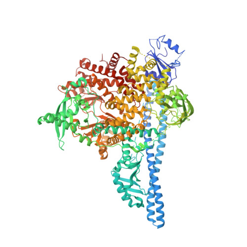
    In the search of PI3K p110α wild type and H1047R mutant selective small molecule leads, an encoded library technology (ELT) campaign against the desired target proteins was performed which led to the discovery of a selective chemotype for PI3K isoforms from a three-cycle DNA encoded library. An X-ray crystal structure of a representative inhibitor from this chemotype demonstrated a unique binding mode in the p110α protein.


  • Organizational Affiliation

    MDR (Molecular Discovery Research) Boston, Platform Technology and Science, GlaxoSmithKline , 830 Winter Street, Waltham, Massachusetts 02451, United States.


Macromolecules
Find similar proteins by:  (by identity cutoff)  |  3D Structure
Entity ID: 1
MoleculeChains Sequence LengthOrganismDetailsImage
Phosphatidylinositol 3-kinase regulatory subunit alpha,Phosphatidylinositol 4,5-bisphosphate 3-kinase catalytic subunit alpha isoform fusion protein1,383Homo sapiensMutation(s): 0 
Gene Names: PIK3R1GRB1PIK3CA
EC: 2.7.1.153 (PDB Primary Data), 2.7.11.1 (PDB Primary Data)
UniProt & NIH Common Fund Data Resources
Find proteins for P27986 (Homo sapiens)
Explore P27986 
Go to UniProtKB:  P27986
PHAROS:  P27986
GTEx:  ENSG00000145675 
Find proteins for P42336 (Homo sapiens)
Explore P42336 
Go to UniProtKB:  P42336
PHAROS:  P42336
GTEx:  ENSG00000121879 
Entity Groups  
Sequence Clusters30% Identity50% Identity70% Identity90% Identity95% Identity100% Identity
UniProt GroupsP42336P27986
Sequence Annotations
Expand
  • Reference Sequence
Small Molecules
Ligands 1 Unique
IDChains Name / Formula / InChI Key2D Diagram3D Interactions
4EL
Query on 4EL

Download Ideal Coordinates CCD File 
B [auth A],
C [auth A],
D [auth A]
3-(6-methoxypyridin-3-yl)-5-[({4-[(5-methyl-1,3,4-thiadiazol-2-yl)sulfamoyl]phenyl}amino)methyl]benzoic acid
C23 H21 N5 O5 S2
CBZPCOUPBNLTKW-UHFFFAOYSA-N
Binding Affinity Annotations 
IDSourceBinding Affinity
4EL BindingDB:  4YKN IC50: min: 8.5, max: 10 (nM) from 2 assay(s)
Experimental Data & Validation

Experimental Data

  • Method: X-RAY DIFFRACTION
  • Resolution: 2.90 Å
  • R-Value Free: 0.247 
  • R-Value Work: 0.202 
  • R-Value Observed: 0.204 
  • Space Group: P 21 21 21
Unit Cell:
Length ( Å )Angle ( ˚ )
a = 84.667α = 90
b = 118.191β = 90
c = 190.281γ = 90
Software Package:
Software NamePurpose
PHENIXrefinement
HKL-2000data scaling
PHASERphasing
SCALAdata scaling
Cootmodel building

Structure Validation

View Full Validation Report


Ligand Structure Quality Assessment 


Entry History 

Deposition Data

  • Released Date: 2015-06-17 
  • Deposition Author(s): Elkins, P.A.

Revision History  (Full details and data files)

  • Version 1.0: 2015-06-17
    Type: Initial release
  • Version 1.1: 2017-11-22
    Changes: Database references, Derived calculations, Refinement description, Source and taxonomy
  • Version 1.2: 2024-02-28
    Changes: Data collection, Database references