4YIZ

Crystal structure of engineered TgAMA1 lacking the DII loop in complex with an Eimeria tenella RON2D3 peptide


Experimental Data Snapshot

  • Method: X-RAY DIFFRACTION
  • Resolution: 2.20 Å
  • R-Value Free: 0.207 
  • R-Value Work: 0.187 
  • R-Value Observed: 0.188 

wwPDB Validation   3D Report Full Report


Ligand Structure Quality Assessment 


This is version 2.1 of the entry. See complete history


Literature

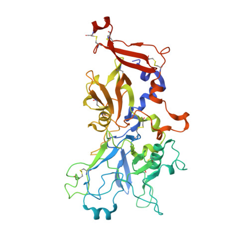
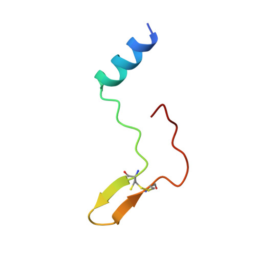
An Extended Surface Loop on Toxoplasma gondii Apical Membrane Antigen 1 (AMA1) Governs Ligand Binding Selectivity.

Parker, M.L.Boulanger, M.J.

(2015) PLoS One 10: e0126206-e0126206

  • DOI: https://doi.org/10.1371/journal.pone.0126206
  • Primary Citation of Related Structures:  
    4YIV, 4YIZ

  • PubMed Abstract: 

    Apicomplexan parasites are the causative agents of globally prevalent diseases including malaria and toxoplasmosis. These obligate intracellular pathogens have evolved a sophisticated host cell invasion strategy that relies on a parasite-host cell junction anchored by interactions between apical membrane antigens (AMAs) on the parasite surface and rhoptry neck 2 (RON2) proteins discharged from the parasite and embedded in the host cell membrane. Key to formation of the AMA1-RON2 complex is displacement of an extended surface loop on AMA1 called the DII loop. While conformational flexibility of the DII loop is required to expose the mature RON2 binding groove, a definitive role of this substructure has not been elucidated. To establish a role of the DII loop in Toxoplasma gondii AMA1, we engineered a form of the protein where the mobile portion of the loop was replaced with a short Gly-Ser linker (TgAMA1ΔDIIloop). Isothermal titration calorimetry measurements with a panel of RON2 peptides revealed an influential role for the DII loop in governing selectivity. Most notably, an Eimeria tenella RON2 (EtRON2) peptide that showed only weak binding to TgAMA1 bound with high affinity to TgAMA1ΔDIIloop. To define the molecular basis for the differential binding, we determined the crystal structure of TgAMA1ΔDIIloop in complex with the EtRON2 peptide. When analyzed in the context of existing AMA1-RON2 structures, spatially distinct anchor points in the AMA1 groove were identified that, when engaged, appear to provide the necessary traction to outcompete the DII loop. Collectively, these data support a model where the AMA1 DII loop serves as a structural gatekeeper to selectively filter out ligands otherwise capable of binding with high affinity in the AMA1 apical groove. These data also highlight the importance of considering the functional implications of the DII loop in the ongoing development of therapeutic intervention strategies targeting the AMA1-RON2 invasion complex.


  • Organizational Affiliation

    Department of Biochemistry & Microbiology, University of Victoria, PO Box 3055 STN CSC, Victoria, BC, V8W 3P6, Canada.


Macromolecules
Find similar proteins by:  (by identity cutoff)  |  3D Structure
Entity ID: 1
MoleculeChains Sequence LengthOrganismDetailsImage
Apical membrane antigen AMA1A,
B [auth C],
C [auth E]
419Toxoplasma gondii ME49Mutation(s): 0 
Gene Names: TGME49_255260
UniProt
Find proteins for S8GKS3 (Toxoplasma gondii (strain ATCC 50611 / Me49))
Explore S8GKS3 
Go to UniProtKB:  S8GKS3
Entity Groups  
Sequence Clusters30% Identity50% Identity70% Identity90% Identity95% Identity100% Identity
UniProt GroupS8GKS3
Sequence Annotations
Expand
  • Reference Sequence
Find similar proteins by:  (by identity cutoff)  |  3D Structure
Entity ID: 2
MoleculeChains Sequence LengthOrganismDetailsImage
Rhoptry neck protein 2, putativeD [auth B],
E [auth D],
F
40Eimeria tenellaMutation(s): 0 
Gene Names: ETH_00012760
UniProt
Find proteins for U6KQJ2 (Eimeria tenella)
Explore U6KQJ2 
Go to UniProtKB:  U6KQJ2
Entity Groups  
Sequence Clusters30% Identity50% Identity70% Identity90% Identity95% Identity100% Identity
UniProt GroupU6KQJ2
Sequence Annotations
Expand
  • Reference Sequence
Small Molecules
Ligands 3 Unique
IDChains Name / Formula / InChI Key2D Diagram3D Interactions
A2G
Query on A2G

Download Ideal Coordinates CCD File 
H [auth A]2-acetamido-2-deoxy-alpha-D-galactopyranose
C8 H15 N O6
OVRNDRQMDRJTHS-CBQIKETKSA-N
NAG
Query on NAG

Download Ideal Coordinates CCD File 
G [auth A],
K [auth C],
N [auth E]
2-acetamido-2-deoxy-beta-D-glucopyranose
C8 H15 N O6
OVRNDRQMDRJTHS-FMDGEEDCSA-N
SO4
Query on SO4

Download Ideal Coordinates CCD File 
I [auth A],
J [auth A],
L [auth C],
M [auth C],
O [auth E]
SULFATE ION
O4 S
QAOWNCQODCNURD-UHFFFAOYSA-L
Experimental Data & Validation

Experimental Data

  • Method: X-RAY DIFFRACTION
  • Resolution: 2.20 Å
  • R-Value Free: 0.207 
  • R-Value Work: 0.187 
  • R-Value Observed: 0.188 
  • Space Group: P 32 2 1
Unit Cell:
Length ( Å )Angle ( ˚ )
a = 265.928α = 90
b = 265.928β = 90
c = 94.16γ = 120
Software Package:
Software NamePurpose
PHENIXrefinement
Blu-Icedata collection
MOSFLMdata reduction
SCALAdata scaling
PHASERphasing
Cootmodel building
PDB_EXTRACTdata extraction

Structure Validation

View Full Validation Report


Ligand Structure Quality Assessment 


Entry History & Funding Information

Deposition Data


Funding OrganizationLocationGrant Number
Canadian Institutes of Health Research (CIHR)CanadaMOP82915

Revision History  (Full details and data files)

  • Version 1.0: 2015-05-20
    Type: Initial release
  • Version 1.1: 2017-09-20
    Changes: Author supporting evidence, Derived calculations, Source and taxonomy
  • Version 1.2: 2020-01-08
    Changes: Author supporting evidence, Data collection
  • Version 2.0: 2020-07-29
    Type: Remediation
    Reason: Carbohydrate remediation
    Changes: Atomic model, Data collection, Derived calculations, Structure summary
  • Version 2.1: 2023-09-27
    Changes: Data collection, Database references, Refinement description, Structure summary