Sperm Lysozyme-Like Protein 1 (SLLP1), an intra-acrosomal oolemmal-binding sperm protein, reveals filamentous organization in protein crystal form.
Zheng, H., Mandal, A., Shumilin, I.A., Chordia, M.D., Panneerdoss, S., Herr, J.C., Minor, W.(2015) Andrology 3: 756-771
- PubMed: 26198801
- DOI: https://doi.org/10.1111/andr.12057
- Primary Citation of Related Structures:
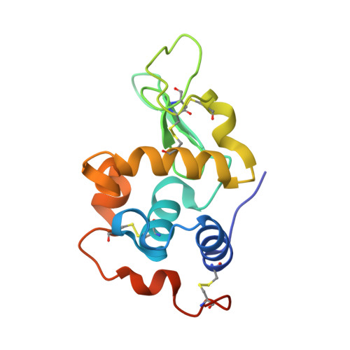
4YF2 - PubMed Abstract:
Sperm lysozyme-like protein 1 (SLLP1) is one of the lysozyme-like proteins predominantly expressed in mammalian testes that lacks bacteriolytic activity, localizes in the sperm acrosome, and exhibits high affinity for an oolemmal receptor, SAS1B. The crystal structure of mouse SLLP1 (mSLLP1) was determined at 2.15 Å resolution. mSLLP1 monomer adopts a structural fold similar to that of chicken/mouse lysozymes retaining all four canonical disulfide bonds. mSLLP1 is distinct from c-lysozyme by substituting two essential catalytic residues (E35T/D52N), exhibiting different surface charge distribution, and by forming helical filaments approximately 75 Å in diameter with a 25 Å central pore comprised of six monomers per helix turn repeating every 33 Å. Cross-species alignment of all reported SLLP1 sequences revealed a set of invariant surface regions comprising a characteristic fingerprint uniquely identifying SLLP1 from other c-lysozyme family members. The fingerprint surface regions reside around the lips of the putative glycan-binding groove including three polar residues (Y33/E46/H113). A flexible salt bridge (E46-R61) was observed covering the glycan-binding groove. The conservation of these regions may be linked to their involvement in oolemmal protein binding. Interaction between SLLP1 monomer and its oolemmal receptor SAS1B was modeled using protein-protein docking algorithms, utilizing the SLLP1 fingerprint regions along with the SAS1B conserved surface regions. This computational model revealed complementarity between the conserved SLLP1/SAS1B interacting surfaces supporting the experimentally observed SLLP1/SAS1B interaction involved in fertilization.
Organizational Affiliation:
Department of Molecular Physiology and Biological Physics, University of Virginia, Charlottesville, VA, USA.