4YDH

The structure of human FMNL1 N-terminal domains bound to Cdc42


Experimental Data Snapshot

  • Method: X-RAY DIFFRACTION
  • Resolution: 3.80 Å
  • R-Value Free: 0.281 
  • R-Value Work: 0.208 
  • R-Value Observed: 0.211 

wwPDB Validation   3D Report Full Report


Ligand Structure Quality Assessment 


This is version 1.2 of the entry. See complete history


Literature

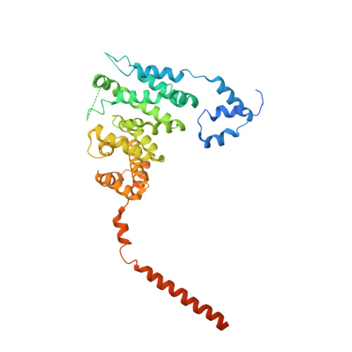
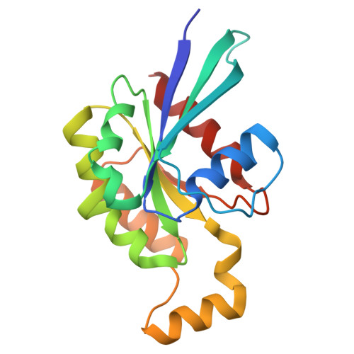
The structure of FMNL2-Cdc42 yields insights into the mechanism of lamellipodia and filopodia formation.

Kuhn, S.Erdmann, C.Kage, F.Block, J.Schwenkmezger, L.Steffen, A.Rottner, K.Geyer, M.

(2015) Nat Commun 6: 7088-7088

  • DOI: https://doi.org/10.1038/ncomms8088
  • Primary Citation of Related Structures:  
    4YC7, 4YDH

  • PubMed Abstract: 

    Formins are actin polymerization factors that elongate unbranched actin filaments at the barbed end. Rho family GTPases activate Diaphanous-related formins through the relief of an autoregulatory interaction. The crystal structures of the N-terminal domains of human FMNL1 and FMNL2 in complex with active Cdc42 show that Cdc42 mediates contacts with all five armadillo repeats of the formin with specific interactions formed by the Rho-GTPase insert helix. Mutation of three residues within Rac1 results in a gain-of-function mutation for FMNL2 binding and reconstitution of the Cdc42 phenotype in vivo. Dimerization of FMNL1 through a parallel coiled coil segment leads to formation of an umbrella-shaped structure that—together with Cdc42—spans more than 15 nm in diameter. The two interacting FMNL-Cdc42 heterodimers expose six membrane interaction motifs on a convex protein surface, the assembly of which may facilitate actin filament elongation at the leading edge of lamellipodia and filopodia.


  • Organizational Affiliation

    1] Center of Advanced European Studies and Research, Group Physical Biochemistry, Ludwig-Erhard-Allee 2, Bonn 53175, Germany [2] Department of Physical Biochemistry, Max Planck Institute of Molecular Physiology, Otto-Hahn-Strasse 11, Dortmund 44227, Germany.


Macromolecules
Find similar proteins by:  (by identity cutoff)  |  3D Structure
Entity ID: 1
MoleculeChains Sequence LengthOrganismDetailsImage
Formin-like protein 1
A, C
433Homo sapiensMutation(s): 0 
Gene Names: FMNL1C17orf1C17orf1BFMNL
UniProt & NIH Common Fund Data Resources
Find proteins for O95466 (Homo sapiens)
Explore O95466 
Go to UniProtKB:  O95466
PHAROS:  O95466
GTEx:  ENSG00000184922 
Entity Groups  
Sequence Clusters30% Identity50% Identity70% Identity90% Identity95% Identity100% Identity
UniProt GroupO95466
Sequence Annotations
Expand
  • Reference Sequence
Find similar proteins by:  (by identity cutoff)  |  3D Structure
Entity ID: 2
MoleculeChains Sequence LengthOrganismDetailsImage
Cell division control protein 42 homolog
B, D
181Homo sapiensMutation(s): 0 
Gene Names: CDC42
UniProt & NIH Common Fund Data Resources
Find proteins for P60953 (Homo sapiens)
Explore P60953 
Go to UniProtKB:  P60953
PHAROS:  P60953
GTEx:  ENSG00000070831 
Entity Groups  
Sequence Clusters30% Identity50% Identity70% Identity90% Identity95% Identity100% Identity
UniProt GroupP60953
Sequence Annotations
Expand
  • Reference Sequence
Experimental Data & Validation

Experimental Data

  • Method: X-RAY DIFFRACTION
  • Resolution: 3.80 Å
  • R-Value Free: 0.281 
  • R-Value Work: 0.208 
  • R-Value Observed: 0.211 
  • Space Group: P 21 21 21
Unit Cell:
Length ( Å )Angle ( ˚ )
a = 87.6α = 90
b = 102.1β = 90
c = 170.6γ = 90
Software Package:
Software NamePurpose
PHENIXrefinement
XDSdata reduction
PHASERphasing
XSCALEdata scaling

Structure Validation

View Full Validation Report


Ligand Structure Quality Assessment 


Entry History 

Deposition Data

Revision History  (Full details and data files)

  • Version 1.0: 2015-05-13
    Type: Initial release
  • Version 1.1: 2019-02-20
    Changes: Advisory, Data collection, Derived calculations
  • Version 1.2: 2024-01-10
    Changes: Data collection, Database references, Derived calculations, Refinement description