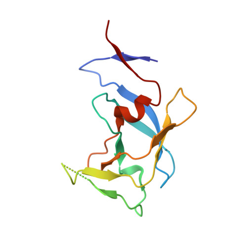
4YDG

Crystal structure of compound 10 in complex with HTLV-1 Protease


Experimental Data Snapshot

  • Method: X-RAY DIFFRACTION
  • Resolution: 3.25 Å
  • R-Value Free: 0.317 
  • R-Value Work: 0.279 
  • R-Value Observed: 0.281 

wwPDB Validation   3D Report Full Report


Ligand Structure Quality Assessment 


This is version 1.1 of the entry. See complete history


Literature

Privileged Structures Meet Human T-Cell Leukemia Virus-1 (HTLV-1): C2-Symmetric 3,4-Disubstituted Pyrrolidines as Nonpeptidic HTLV-1 Protease Inhibitors.

Kuhnert, M.Blum, A.Steuber, H.Diederich, W.E.

(2015) J Med Chem 58: 4845-4850

  • DOI: https://doi.org/10.1021/acs.jmedchem.5b00346
  • Primary Citation of Related Structures:  
    4YDF, 4YDG

  • PubMed Abstract: 

    3,4-disubstituted pyrrolidines originally designed to inhibit the closely related HIV-1 protease were evaluated as privileged structures against HTLV-1 protease (HTLV-1 PR). The most potent inhibitor of this series exhibits two-digit nanomolar affinity and represents, to the best of our knowledge, the most potent nonpeptidic inhibitor of HTLV-1 PR described so far. The X-ray structures of two representatives bound to HTLV-1 PR were determined, and the structural basis of their affinity is discussed.


  • Organizational Affiliation

    †Institut für Pharmazeutische Chemie, Philipps-Universität Marburg, Hans-Meerwein-Straße 3, 35043 Marburg, Germany.


Macromolecules
Find similar proteins by:  (by identity cutoff)  |  3D Structure
Entity ID: 1
MoleculeChains Sequence LengthOrganismDetailsImage
HTLV-1 protease
A, B
116Human T-cell lymphotrophic virus type 1 (strain ATK)Mutation(s): 1 
Gene Names: gag-pro-pol
EC: 3.4.23 (PDB Primary Data), 2.7.7.49 (PDB Primary Data), 2.7.7.7 (PDB Primary Data), 3.1.26.4 (PDB Primary Data)
UniProt
Find proteins for P03362 (Human T-cell leukemia virus 1 (strain Japan ATK-1 subtype A))
Explore P03362 
Go to UniProtKB:  P03362
Entity Groups  
Sequence Clusters30% Identity50% Identity70% Identity90% Identity95% Identity100% Identity
UniProt GroupP03362
Sequence Annotations
Expand
  • Reference Sequence
Small Molecules
Experimental Data & Validation

Experimental Data

  • Method: X-RAY DIFFRACTION
  • Resolution: 3.25 Å
  • R-Value Free: 0.317 
  • R-Value Work: 0.279 
  • R-Value Observed: 0.281 
  • Space Group: P 63 2 2
Unit Cell:
Length ( Å )Angle ( ˚ )
a = 77.61α = 90
b = 77.61β = 90
c = 160.23γ = 120
Software Package:
Software NamePurpose
PHENIXrefinement
XDSdata reduction
XSCALEdata scaling
PHASERphasing

Structure Validation

View Full Validation Report


Ligand Structure Quality Assessment 


Entry History 

Deposition Data

Revision History  (Full details and data files)

  • Version 1.0: 2016-02-03
    Type: Initial release
  • Version 1.1: 2024-01-10
    Changes: Data collection, Database references, Refinement description