4Y5F

PAS-GAF fragment from Deinococcus radiodurans BphP assembled with BV - Y307S, high dose


Experimental Data Snapshot

  • Method: X-RAY DIFFRACTION
  • Resolution: 1.70 Å
  • R-Value Free: 0.214 
  • R-Value Work: 0.143 
  • R-Value Observed: 0.146 

wwPDB Validation   3D Report Full Report


Ligand Structure Quality Assessment 


This is version 1.4 of the entry. See complete history


Literature

X-ray radiation induces deprotonation of the bilin chromophore in crystalline D. radiodurans phytochrome.

Li, F.Burgie, E.S.Yu, T.Heroux, A.Schatz, G.C.Vierstra, R.D.Orville, A.M.

(2015) J Am Chem Soc 137: 2792-2795

  • DOI: https://doi.org/10.1021/ja510923m
  • Primary Citation of Related Structures:  
    4Y3I, 4Y5F

  • PubMed Abstract: 

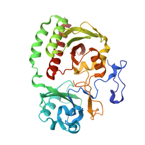
    We report that in the red light-absorbing (Pr) state, the bilin chromophore of the Deinococcus radiodurans proteobacterial phytochrome (DrBphP) is hypersensitive to X-ray photons used in typical synchrotron X-ray protein crystallography experiments. This causes the otherwise fully protonated chromophore to deprotonate without additional major structural changes. These results have major implications for our understanding of the structural and chemical characteristics of the resting and intermediate states of phytochromes and other photoreceptor proteins.


  • Organizational Affiliation

    Photon Sciences Directorate and ∥Biosciences Department, Brookhaven National Laboratory , Upton, New York 11973, United States.


Macromolecules
Find similar proteins by:  (by identity cutoff)  |  3D Structure
Entity ID: 1
MoleculeChains Sequence LengthOrganismDetailsImage
Bacteriophytochrome319Deinococcus radiodurans R1 = ATCC 13939 = DSM 20539Mutation(s): 1 
Gene Names: bphPDR_A0050
EC: 2.7.13.3
UniProt
Find proteins for Q9RZA4 (Deinococcus radiodurans (strain ATCC 13939 / DSM 20539 / JCM 16871 / CCUG 27074 / LMG 4051 / NBRC 15346 / NCIMB 9279 / VKM B-1422 / R1))
Explore Q9RZA4 
Go to UniProtKB:  Q9RZA4
Entity Groups  
Sequence Clusters30% Identity50% Identity70% Identity90% Identity95% Identity100% Identity
UniProt GroupQ9RZA4
Sequence Annotations
Expand
  • Reference Sequence
Small Molecules
Ligands 1 Unique
IDChains Name / Formula / InChI Key2D Diagram3D Interactions
LBV
Query on LBV

Download Ideal Coordinates CCD File 
B [auth A]3-[2-[(Z)-[3-(2-carboxyethyl)-5-[(Z)-(4-ethenyl-3-methyl-5-oxidanylidene-pyrrol-2-ylidene)methyl]-4-methyl-pyrrol-1-ium -2-ylidene]methyl]-5-[(Z)-[(3E)-3-ethylidene-4-methyl-5-oxidanylidene-pyrrolidin-2-ylidene]methyl]-4-methyl-1H-pyrrol-3- yl]propanoic acid
C33 H37 N4 O6
DKMLMZVDTGOEGU-ISEYCTJISA-O
Experimental Data & Validation

Experimental Data

  • Method: X-RAY DIFFRACTION
  • Resolution: 1.70 Å
  • R-Value Free: 0.214 
  • R-Value Work: 0.143 
  • R-Value Observed: 0.146 
  • Space Group: C 1 2 1
Unit Cell:
Length ( Å )Angle ( ˚ )
a = 89.274α = 90
b = 51.803β = 116.22
c = 80.272γ = 90
Software Package:
Software NamePurpose
REFMACrefinement
HKL-2000data reduction
HKL-2000data scaling

Structure Validation

View Full Validation Report


Ligand Structure Quality Assessment 


Entry History & Funding Information

Deposition Data


Funding OrganizationLocationGrant Number
National Institutes of Health/National Institute of General Medical Sciences (NIH/NIGMS)United States8P41GM103473-16
Department of Energy (DOE, United States)United StatesFWP BO-70
National Science Foundation (NSF, United States)United StatesMCB-1329956
National Science Foundation (NSF, United States)United StatesCHE-1147335
Department of Energy (DOE, United States)United StatesDE-AC02-98CH10886

Revision History  (Full details and data files)

  • Version 1.0: 2015-05-20
    Type: Initial release
  • Version 1.1: 2017-09-06
    Changes: Author supporting evidence, Derived calculations, Source and taxonomy
  • Version 1.2: 2017-11-22
    Changes: Refinement description
  • Version 1.3: 2019-11-27
    Changes: Author supporting evidence
  • Version 1.4: 2023-09-27
    Changes: Data collection, Database references, Derived calculations, Refinement description, Structure summary