4Y46

Pyridopyrimidinone Derivatives as Potent and Selective c-Jun N-Terminal Kinase (JNK) inhibitors


Experimental Data Snapshot

  • Method: X-RAY DIFFRACTION
  • Resolution: 2.04 Å
  • R-Value Free: 0.265 
  • R-Value Work: 0.212 
  • R-Value Observed: 0.214 

wwPDB Validation   3D Report Full Report


Ligand Structure Quality Assessment 


This is version 1.1 of the entry. See complete history


Literature

Pyridopyrimidinone Derivatives as Potent and Selective c-Jun N-Terminal Kinase (JNK) Inhibitors.

Zheng, K.Park, C.M.Iqbal, S.Hernandez, P.Park, H.LoGrasso, P.V.Feng, Y.

(2015) ACS Med Chem Lett 6: 413-418

  • DOI: https://doi.org/10.1021/ml500474d
  • Primary Citation of Related Structures:  
    4Y46, 4Y5H

  • PubMed Abstract: 

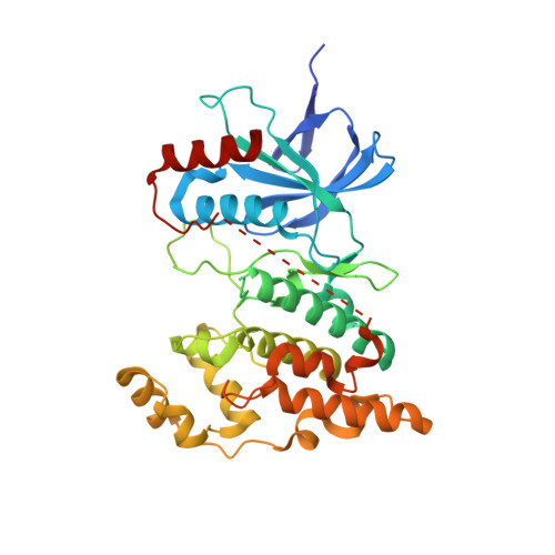
    A novel series of 2-aminopyridopyrimidinone based JNK (c-jun N-terminal kinase) inhibitors were discovered and developed. Structure-activity relationships (SARs) were systematically developed utilizing biochemical and cell based assays and in vitro and in vivo drug metabolism and pharmacokinetic (DMPK) studies. Through the optimization of lead compound 1, several potent and selective JNK inhibitors with high oral bioavailability were developed. Inhibitor 13 was a potent JNK3 inhibitor (IC50 = 15 nM), had high selectivity against p38 (IC50 > 10 μM), had high potency in functional cell based assays, and had high stability in human liver microsome (t 1/2 = 76 min), a clean CYP-450 inhibition profile, and excellent oral bioavailability (%F = 87). Moreover, cocrystal structures of compounds 13 and 22 in JNK3 were solved at 2.0 Å. These structures elucidated the binding mode (Type-I binding) and can pave the way for further inhibitor design of this pyridopyrimidinone scaffold for JNK inhibition.


  • Organizational Affiliation

    Medicinal Chemistry, Discovery Biology, Crystallography/Modeling Core Facility, Translational Research Institute, and Department of Molecular Therapeutics, The Scripps Research Institute , 130 Scripps Way, #2A1, Jupiter, Florida 33458, United States.


Macromolecules
Find similar proteins by:  (by identity cutoff)  |  3D Structure
Entity ID: 1
MoleculeChains Sequence LengthOrganismDetailsImage
Mitogen-activated protein kinase 10366Homo sapiensMutation(s): 0 
Gene Names: MAPK10JNK3JNK3APRKM10SAPK1B
EC: 2.7.11.24
UniProt & NIH Common Fund Data Resources
Find proteins for P53779 (Homo sapiens)
Explore P53779 
Go to UniProtKB:  P53779
PHAROS:  P53779
GTEx:  ENSG00000109339 
Entity Groups  
Sequence Clusters30% Identity50% Identity70% Identity90% Identity95% Identity100% Identity
UniProt GroupP53779
Sequence Annotations
Expand
  • Reference Sequence
Small Molecules
Ligands 1 Unique
IDChains Name / Formula / InChI Key2D Diagram3D Interactions
4F2
Query on 4F2

Download Ideal Coordinates CCD File 
B [auth A]1-{trans-4-[(8-cyclopentyl-7-oxo-7,8-dihydropyrido[2,3-d]pyrimidin-2-yl)amino]cyclohexyl}-3-propan-2-ylurea
C22 H32 N6 O2
QJPUIIKZKMYPSK-QAQDUYKDSA-N
Binding Affinity Annotations 
IDSourceBinding Affinity
4F2 Binding MOAD:  4Y46 IC50: 11 (nM) from 1 assay(s)
BindingDB:  4Y46 IC50: min: 7, max: 12 (nM) from 3 assay(s)
Experimental Data & Validation

Experimental Data

  • Method: X-RAY DIFFRACTION
  • Resolution: 2.04 Å
  • R-Value Free: 0.265 
  • R-Value Work: 0.212 
  • R-Value Observed: 0.214 
  • Space Group: P 21 21 21
Unit Cell:
Length ( Å )Angle ( ˚ )
a = 53.8α = 90
b = 72.14β = 90
c = 107.94γ = 90
Software Package:
Software NamePurpose
PHENIXrefinement
HKL-2000data reduction
HKL-2000data scaling
PHASERphasing

Structure Validation

View Full Validation Report


Ligand Structure Quality Assessment 


Entry History 

Deposition Data

  • Released Date: 2015-05-06 
  • Deposition Author(s): Park, H.

Revision History  (Full details and data files)

  • Version 1.0: 2015-05-06
    Type: Initial release
  • Version 1.1: 2024-02-28
    Changes: Data collection, Database references, Derived calculations, Source and taxonomy