4XWH

Crystal structure of the human N-acetyl-alpha-glucosaminidase


Experimental Data Snapshot

  • Method: X-RAY DIFFRACTION
  • Resolution: 2.32 Å
  • R-Value Free: 0.186 
  • R-Value Work: 0.166 
  • R-Value Observed: 0.167 

wwPDB Validation   3D Report Full Report


Ligand Structure Quality Assessment 


This is version 2.1 of the entry. See complete history


Literature

Structural characterization of the alpha-N-acetylglucosaminidase, a key enzyme in the pathogenesis of Sanfilippo syndrome B.

Birrane, G.Dassier, A.L.Romashko, A.Lundberg, D.Holmes, K.Cottle, T.Norton, A.W.Zhang, B.Concino, M.F.Meiyappan, M.

(2019) J Struct Biol 205: 65-71

  • DOI: https://doi.org/10.1016/j.jsb.2019.02.005
  • Primary Citation of Related Structures:  
    4XWH

  • PubMed Abstract: 

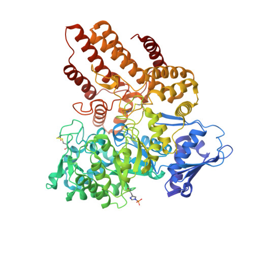
    Mucopolysaccharidosis III B (MPS III-B) is a rare lysosomal storage disorder caused by deficiencies in Alpha-N-acetylglucosaminidase (NAGLU) for which there is currently no cure, and present treatment is largely supportive. Understanding the structure of NAGLU may allow for identification of novel therapeutic targets for MPS III-B. Here we describe the first crystal structure of human NAGLU, determined to a resolution of 2.3 Å. The crystal structure reveals a novel homotrimeric configuration, maintained primarily by hydrophobic and electrostatic interactions via domain II of three contiguous domains from the N- to C-terminus. The active site cleft is located between domains II and III. Catalytic glutamate residues, E316 and E446, are located at the top of the (α/β) 8 barrel structure in domain II. We utilized the three-dimensional structure of NAGLU to map several MPS III-B mutations, and hypothesize their functional consequences. Revealing atomic level structural information about this critical lysosomal enzyme paves the way for the design of novel therapeutics to target the underlying causes of MPS III-B.


  • Organizational Affiliation

    Division of Experimental Medicine, Department of Medicine, Beth Israel Deaconess Medical Center, 99 Brookline Ave., Boston, MA 02215, United States.


Macromolecules
Find similar proteins by:  (by identity cutoff)  |  3D Structure
Entity ID: 1
MoleculeChains Sequence LengthOrganismDetailsImage
Alpha-N-acetylglucosaminidase720Homo sapiensMutation(s): 0 
Gene Names: NAGLUUFHSD1
EC: 3.2.1.50
UniProt & NIH Common Fund Data Resources
Find proteins for P54802 (Homo sapiens)
Explore P54802 
Go to UniProtKB:  P54802
PHAROS:  P54802
GTEx:  ENSG00000108784 
Entity Groups  
Sequence Clusters30% Identity50% Identity70% Identity90% Identity95% Identity100% Identity
UniProt GroupP54802
Sequence Annotations
Expand
  • Reference Sequence
Oligosaccharides

Help

Entity ID: 2
MoleculeChains Length2D Diagram Glycosylation3D Interactions
beta-D-mannopyranose-(1-4)-2-acetamido-2-deoxy-beta-D-glucopyranose-(1-4)-2-acetamido-2-deoxy-beta-D-glucopyranose
B
3N-Glycosylation
Glycosylation Resources
GlyTouCan:  G15407YE
GlyCosmos:  G15407YE
GlyGen:  G15407YE
Entity ID: 3
MoleculeChains Length2D Diagram Glycosylation3D Interactions
2-acetamido-2-deoxy-beta-D-glucopyranose-(1-4)-2-acetamido-2-deoxy-beta-D-glucopyranose
C, D
2N-Glycosylation
Glycosylation Resources
GlyTouCan:  G42666HT
GlyCosmos:  G42666HT
GlyGen:  G42666HT
Small Molecules
Ligands 3 Unique
IDChains Name / Formula / InChI Key2D Diagram3D Interactions
NAG
Query on NAG

Download Ideal Coordinates CCD File 
E [auth A],
F [auth A],
G [auth A]
2-acetamido-2-deoxy-beta-D-glucopyranose
C8 H15 N O6
OVRNDRQMDRJTHS-FMDGEEDCSA-N
XYL
Query on XYL

Download Ideal Coordinates CCD File 
K [auth A],
L [auth A]
Xylitol
C5 H12 O5
HEBKCHPVOIAQTA-SCDXWVJYSA-N
GOL
Query on GOL

Download Ideal Coordinates CCD File 
H [auth A],
I [auth A],
J [auth A]
GLYCEROL
C3 H8 O3
PEDCQBHIVMGVHV-UHFFFAOYSA-N
Modified Residues  1 Unique
IDChains TypeFormula2D DiagramParent
NEP
Query on NEP
A
L-PEPTIDE LINKINGC6 H10 N3 O5 PHIS
Experimental Data & Validation

Experimental Data

  • Method: X-RAY DIFFRACTION
  • Resolution: 2.32 Å
  • R-Value Free: 0.186 
  • R-Value Work: 0.166 
  • R-Value Observed: 0.167 
  • Space Group: P 63
Unit Cell:
Length ( Å )Angle ( ˚ )
a = 205.088α = 90
b = 205.088β = 90
c = 78.398γ = 120
Software Package:
Software NamePurpose
REFMACrefinement
CBASSdata collection
HKL-2000data scaling
PHASERphasing
HKL-2000data reduction

Structure Validation

View Full Validation Report


Ligand Structure Quality Assessment 


Entry History 

Deposition Data

Revision History  (Full details and data files)

  • Version 1.0: 2016-02-03
    Type: Initial release
  • Version 1.1: 2017-11-22
    Changes: Derived calculations, Refinement description
  • Version 1.2: 2019-07-17
    Changes: Data collection, Database references
  • Version 2.0: 2020-07-29
    Type: Remediation
    Reason: Carbohydrate remediation
    Changes: Atomic model, Data collection, Derived calculations, Structure summary
  • Version 2.1: 2023-09-27
    Changes: Data collection, Database references, Refinement description, Structure summary