


Crystal structure of Dihydroorotate Dehydrogenase form Mycobacterium Tuberculosis
Kishor, C., Addlagatta, A.To be published.
Experimental Data Snapshot
Entity ID: 1 | |||||
|---|---|---|---|---|---|
| Molecule | Chains | Sequence Length | Organism | Details | Image |
| Dihydroorotate dehydrogenase (quinone) | 344 | Mycobacterium tuberculosis H37Ra | Mutation(s): 0 Gene Names: pyrD, MRA_2153 EC: 1.3.5.2 | 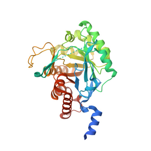 | |
UniProt | |||||
Find proteins for P9WHL1 (Mycobacterium tuberculosis (strain ATCC 25618 / H37Rv)) Explore P9WHL1 Go to UniProtKB: P9WHL1 | |||||
Entity Groups | |||||
| Sequence Clusters | 30% Identity50% Identity70% Identity90% Identity95% Identity100% Identity | ||||
| UniProt Group | P9WHL1 | ||||
Sequence AnnotationsExpand | |||||
| |||||
| Ligands 3 Unique | |||||
|---|---|---|---|---|---|
| ID | Chains | Name / Formula / InChI Key | 2D Diagram | 3D Interactions | |
| FMN Query on FMN | C [auth A], G [auth B] | FLAVIN MONONUCLEOTIDE C17 H21 N4 O9 P FVTCRASFADXXNN-SCRDCRAPSA-N |  | ||
| CL Query on CL | D [auth A], E [auth A], H [auth B], I [auth B], J [auth B] | CHLORIDE ION Cl VEXZGXHMUGYJMC-UHFFFAOYSA-M |  | ||
| MG Query on MG | F [auth B] | MAGNESIUM ION Mg JLVVSXFLKOJNIY-UHFFFAOYSA-N |  | ||
| Length ( Å ) | Angle ( ˚ ) |
|---|---|
| a = 48.335 | α = 90 |
| b = 106.921 | β = 106.21 |
| c = 65.089 | γ = 90 |
| Software Name | Purpose |
|---|---|
| REFMAC | refinement |
| HKL-2000 | data reduction |
| DENZO | data scaling |
| MOLREP | model building |
RCSB PDB (citation) is hosted by
RCSB PDB is a member of the

