4XNW

The human P2Y1 receptor in complex with MRS2500


Experimental Data Snapshot

  • Method: X-RAY DIFFRACTION
  • Resolution: 2.70 Å
  • R-Value Free: 0.267 
  • R-Value Work: 0.218 
  • R-Value Observed: 0.221 

wwPDB Validation   3D Report Full Report


Ligand Structure Quality Assessment 


This is version 1.3 of the entry. See complete history


Literature

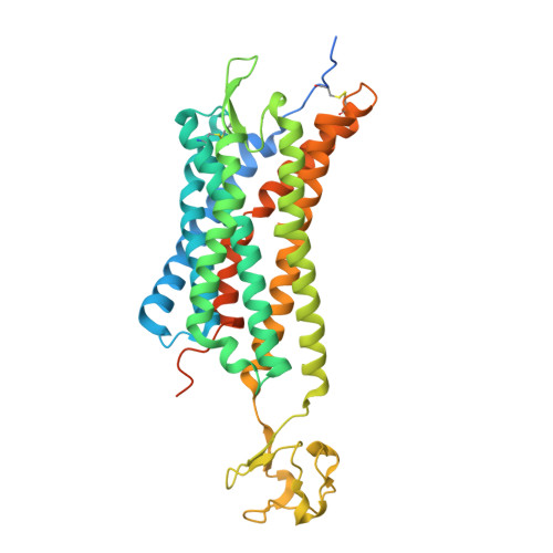
Two disparate ligand-binding sites in the human P2Y1 receptor

Zhang, D.Gao, Z.G.Zhang, K.Kiselev, E.Crane, S.Wang, J.Paoletta, S.Yi, C.Ma, L.Zhang, W.Han, G.W.Liu, H.Cherezov, V.Katritch, V.Jiang, H.Stevens, R.C.Jacobson, K.A.Zhao, Q.Wu, B.

(2015) Nature 520: 317-321

  • DOI: https://doi.org/10.1038/nature14287
  • Primary Citation of Related Structures:  
    4XNV, 4XNW

  • PubMed Abstract: 

    In response to adenosine 5'-diphosphate, the P2Y1 receptor (P2Y1R) facilitates platelet aggregation, and thus serves as an important antithrombotic drug target. Here we report the crystal structures of the human P2Y1R in complex with a nucleotide antagonist MRS2500 at 2.7 Å resolution, and with a non-nucleotide antagonist BPTU at 2.2 Å resolution. The structures reveal two distinct ligand-binding sites, providing atomic details of P2Y1R's unique ligand-binding modes. MRS2500 recognizes a binding site within the seven transmembrane bundle of P2Y1R, which is different in shape and location from the nucleotide binding site in the previously determined structure of P2Y12R, representative of another P2YR subfamily. BPTU binds to an allosteric pocket on the external receptor interface with the lipid bilayer, making it the first structurally characterized selective G-protein-coupled receptor (GPCR) ligand located entirely outside of the helical bundle. These high-resolution insights into P2Y1R should enable discovery of new orthosteric and allosteric antithrombotic drugs with reduced adverse effects.


  • Organizational Affiliation

    CAS Key Laboratory of Receptor Research, Shanghai Institute of Materia Medica, Chinese Academy of Sciences, 555 Zuchongzhi Road, Pudong, Shanghai 201203, China.


Macromolecules
Find similar proteins by:  (by identity cutoff)  |  3D Structure
Entity ID: 1
MoleculeChains Sequence LengthOrganismDetailsImage
P2Y purinoceptor 1,Rubredoxin,P2Y purinoceptor 1A,
B [auth C]
421Homo sapiensClostridium pasteurianumMutation(s): 1 
Gene Names: P2RY1
Membrane Entity: Yes 
UniProt & NIH Common Fund Data Resources
Find proteins for P00268 (Clostridium pasteurianum)
Explore P00268 
Go to UniProtKB:  P00268
Find proteins for P47900 (Homo sapiens)
Explore P47900 
Go to UniProtKB:  P47900
PHAROS:  P47900
GTEx:  ENSG00000169860 
Entity Groups  
Sequence Clusters30% Identity50% Identity70% Identity90% Identity95% Identity100% Identity
UniProt GroupsP00268P47900
Sequence Annotations
Expand
  • Reference Sequence
Small Molecules
Binding Affinity Annotations 
IDSourceBinding Affinity
2ID BindingDB:  4XNW Ki: 0.78 (nM) from 1 assay(s)
IC50: 8.4 (nM) from 1 assay(s)
Experimental Data & Validation

Experimental Data

  • Method: X-RAY DIFFRACTION
  • Resolution: 2.70 Å
  • R-Value Free: 0.267 
  • R-Value Work: 0.218 
  • R-Value Observed: 0.221 
  • Space Group: P 1
Unit Cell:
Length ( Å )Angle ( ˚ )
a = 40.67α = 101.82
b = 64.97β = 95.7
c = 77.57γ = 100.85
Software Package:
Software NamePurpose
REFMACrefinement
HKL-2000data reduction
SCALEPACKdata scaling
PHASERphasing

Structure Validation

View Full Validation Report


Ligand Structure Quality Assessment 


Entry History 

Deposition Data

Revision History  (Full details and data files)

  • Version 1.0: 2015-04-01
    Type: Initial release
  • Version 1.1: 2015-04-22
    Changes: Database references
  • Version 1.2: 2015-04-29
    Changes: Structure summary
  • Version 1.3: 2020-02-05
    Changes: Data collection, Database references, Derived calculations, Source and taxonomy, Structure summary