


Crystal Structure of E. coli Aminopeptidase N in complex with Beta Alanine
Addlagatta, A., Gumpena, R.To be published.
Experimental Data Snapshot
wwPDB Validation 3D Report Full Report

Entity ID: 1 | |||||
|---|---|---|---|---|---|
| Molecule | Chains | Sequence Length | Organism | Details | Image |
| Aminopeptidase N | 866 | Escherichia coli K-12 | Mutation(s): 0 Gene Names: pepN, b0932, JW0915 EC: 3.4.11.2 | 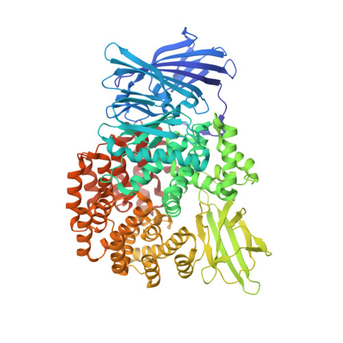 | |
UniProt | |||||
Find proteins for P04825 (Escherichia coli (strain K12)) Explore P04825 Go to UniProtKB: P04825 | |||||
Entity Groups | |||||
| Sequence Clusters | 30% Identity50% Identity70% Identity90% Identity95% Identity100% Identity | ||||
| UniProt Group | P04825 | ||||
Sequence AnnotationsExpand | |||||
| |||||
| Ligands 4 Unique | |||||
|---|---|---|---|---|---|
| ID | Chains | Name / Formula / InChI Key | 2D Diagram | 3D Interactions | |
| MLI Query on MLI | F [auth A], G [auth A] | MALONATE ION C3 H2 O4 OFOBLEOULBTSOW-UHFFFAOYSA-L |  | ||
| BAL Query on BAL | C [auth A] | BETA-ALANINE C3 H7 N O2 UCMIRNVEIXFBKS-UHFFFAOYSA-N |  | ||
| ZN Query on ZN | B [auth A] | ZINC ION Zn PTFCDOFLOPIGGS-UHFFFAOYSA-N |  | ||
| NA Query on NA | D [auth A], E [auth A] | SODIUM ION Na FKNQFGJONOIPTF-UHFFFAOYSA-N |  | ||
| Length ( Å ) | Angle ( ˚ ) |
|---|---|
| a = 120.884 | α = 90 |
| b = 120.884 | β = 90 |
| c = 170.379 | γ = 120 |
| Software Name | Purpose |
|---|---|
| REFMAC | refinement |
| HKL-3000 | data reduction |
| HKL-3000 | data scaling |
| MOLREP | phasing |
RCSB PDB (citation) is hosted by
RCSB PDB is a member of the

