


Crystal Structure of LuxS from Streptococcus suis
Wang, Y., Mao, X., Lu, C.To be published.
Experimental Data Snapshot
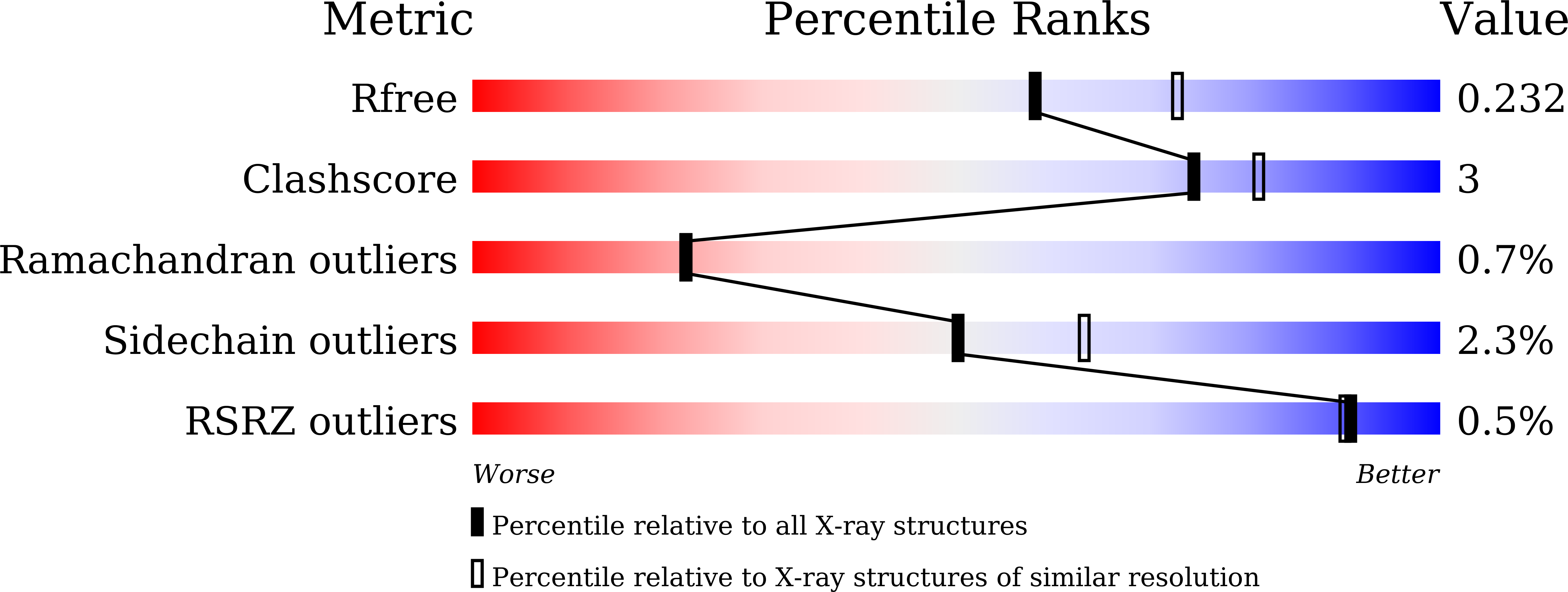
wwPDB Validation 3D Report Full Report
Entity ID: 1 | |||||
|---|---|---|---|---|---|
| Molecule | Chains | Sequence Length | Organism | Details | Image |
| S-ribosylhomocysteine lyase | 194 | Streptococcus suis | Mutation(s): 0 Gene Names: luxS EC: 4.4.1.21 | 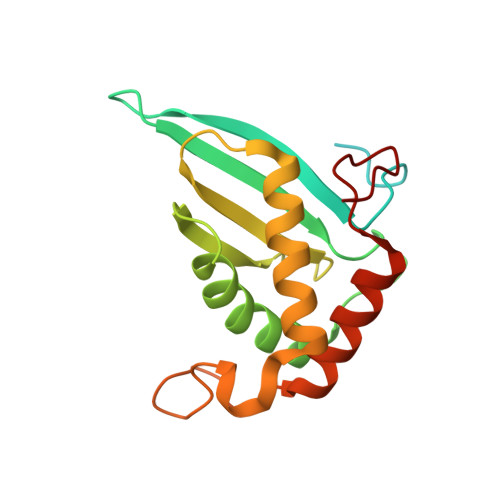 | |
UniProt | |||||
Find proteins for B2CMA5 (Streptococcus suis) Explore B2CMA5 Go to UniProtKB: B2CMA5 | |||||
Entity Groups | |||||
| Sequence Clusters | 30% Identity50% Identity70% Identity90% Identity95% Identity100% Identity | ||||
| UniProt Group | B2CMA5 | ||||
Sequence AnnotationsExpand | |||||
| |||||
| Ligands 1 Unique | |||||
|---|---|---|---|---|---|
| ID | Chains | Name / Formula / InChI Key | 2D Diagram | 3D Interactions | |
| ZN Query on ZN | E [auth A], F [auth B], G [auth C], H [auth D] | ZINC ION Zn PTFCDOFLOPIGGS-UHFFFAOYSA-N |  | ||
| Length ( Å ) | Angle ( ˚ ) |
|---|---|
| a = 74.89 | α = 90 |
| b = 86.25 | β = 90 |
| c = 103.5 | γ = 90 |
| Software Name | Purpose |
|---|---|
| PHENIX | refinement |
| MOSFLM | data processing |
| SCALA | data scaling |
| PHASER | phasing |
RCSB PDB (citation) is hosted by
RCSB PDB is a member of the

