4XBU

In vitro Crystal Structure of PAK4 in complex with Inka peptide


Experimental Data Snapshot

  • Method: X-RAY DIFFRACTION
  • Resolution: 2.06 Å
  • R-Value Free: 0.243 
  • R-Value Work: 0.199 
  • R-Value Observed: 0.201 

wwPDB Validation   3D Report Full Report


This is version 1.3 of the entry. See complete history


Literature

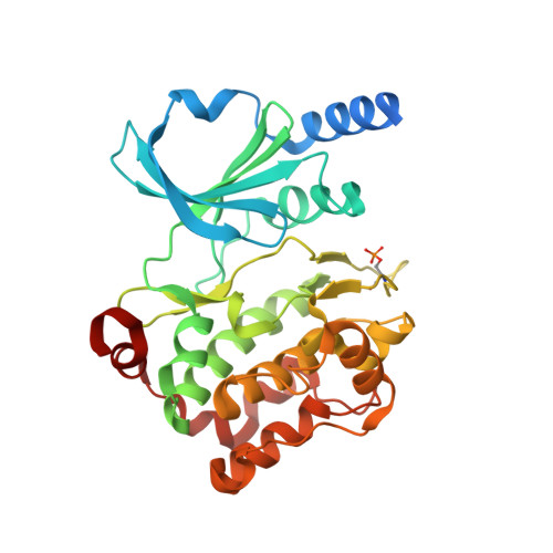
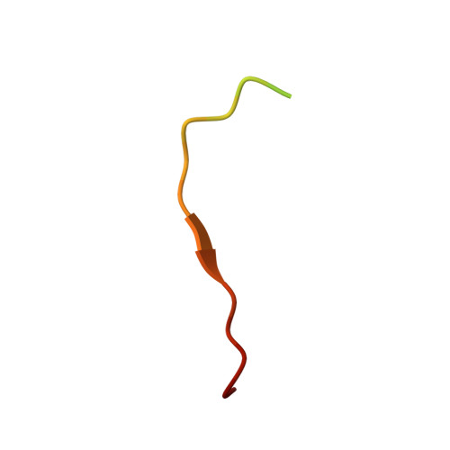
An in cellulo-derived structure of PAK4 in complex with its inhibitor Inka1

Baskaran, Y.Ang, K.C.Anekal, P.V.Chan, W.L.Grimes, J.M.Manser, E.Robinson, R.C.

(2015) Nat Commun 6: 8681-8681

  • DOI: https://doi.org/10.1038/ncomms9681
  • Primary Citation of Related Structures:  
    4XBR, 4XBU

  • PubMed Abstract: 

    PAK4 is a metazoan-specific kinase acting downstream of Cdc42. Here we describe the structure of human PAK4 in complex with Inka1, a potent endogenous kinase inhibitor. Using single mammalian cells containing crystals 50 μm in length, we have determined the in cellulo crystal structure at 2.95 Å resolution, which reveals the details of how the PAK4 catalytic domain binds cellular ATP and the Inka1 inhibitor. The crystal lattice consists only of PAK4-PAK4 contacts, which form a hexagonal array with channels of 80 Å in diameter that run the length of the crystal. The crystal accommodates a variety of other proteins when fused to the kinase inhibitor. Inka1-GFP was used to monitor the process crystal formation in living cells. Similar derivatives of Inka1 will allow us to study the effects of PAK4 inhibition in cells and model organisms, to allow better validation of therapeutic agents targeting PAK4.


  • Organizational Affiliation

    Institute of Molecular and Cell Biology, A*STAR (Agency for Science, Technology and Research), Biopolis, Proteos Building, 61 Biopolis Drive, 8-15, Singapore 138673, Singapore.


Macromolecules
Find similar proteins by:  (by identity cutoff)  |  3D Structure
Entity ID: 1
MoleculeChains Sequence LengthOrganismDetailsImage
Serine/threonine-protein kinase PAK 4326Homo sapiensMutation(s): 0 
Gene Names: PAK4KIAA1142
EC: 2.7.11.1
UniProt & NIH Common Fund Data Resources
Find proteins for O96013 (Homo sapiens)
Explore O96013 
Go to UniProtKB:  O96013
PHAROS:  O96013
GTEx:  ENSG00000130669 
Entity Groups  
Sequence Clusters30% Identity50% Identity70% Identity90% Identity95% Identity100% Identity
UniProt GroupO96013
Sequence Annotations
Expand
  • Reference Sequence

Find similar proteins by:  Sequence   |   3D Structure  

Entity ID: 2
MoleculeChains Sequence LengthOrganismDetailsImage
Protein FAM212A24Homo sapiensMutation(s): 0 
UniProt & NIH Common Fund Data Resources
Find proteins for Q96EL1 (Homo sapiens)
Explore Q96EL1 
Go to UniProtKB:  Q96EL1
PHAROS:  Q96EL1
Entity Groups  
Sequence Clusters30% Identity50% Identity70% Identity90% Identity95% Identity100% Identity
UniProt GroupQ96EL1
Sequence Annotations
Expand
  • Reference Sequence
Small Molecules
Modified Residues  1 Unique
IDChains TypeFormula2D DiagramParent
SEP
Query on SEP
A
L-PEPTIDE LINKINGC3 H8 N O6 PSER
Experimental Data & Validation

Experimental Data

  • Method: X-RAY DIFFRACTION
  • Resolution: 2.06 Å
  • R-Value Free: 0.243 
  • R-Value Work: 0.199 
  • R-Value Observed: 0.201 
  • Space Group: P 41 21 2
Unit Cell:
Length ( Å )Angle ( ˚ )
a = 65.62α = 90
b = 65.62β = 90
c = 184.15γ = 90
Software Package:
Software NamePurpose
REFMACrefinement
xia2data reduction
xia2data scaling
xia2data processing

Structure Validation

View Full Validation Report


Entry History & Funding Information

Deposition Data


Funding OrganizationLocationGrant Number
Agency for Science, Technology and ResearchSingapore--

Revision History  (Full details and data files)

  • Version 1.0: 2015-12-02
    Type: Initial release
  • Version 1.1: 2015-12-09
    Changes: Database references
  • Version 1.2: 2017-10-04
    Changes: Data collection, Derived calculations
  • Version 1.3: 2023-11-08
    Changes: Data collection, Database references, Refinement description