4XB6

Structure of the E. coli C-P lyase core complex


Experimental Data Snapshot

  • Method: X-RAY DIFFRACTION
  • Resolution: 1.70 Å
  • R-Value Free: 0.176 
  • R-Value Work: 0.148 
  • R-Value Observed: 0.150 

wwPDB Validation   3D Report Full Report


This is version 1.3 of the entry. See complete history


Literature

Structural insights into the bacterial carbon-phosphorus lyase machinery.

Seweryn, P.Van, L.B.Kjeldgaard, M.Russo, C.J.Passmore, L.A.Hove-Jensen, B.Jochimsen, B.Brodersen, D.E.

(2015) Nature 525: 68-72

  • DOI: https://doi.org/10.1038/nature14683
  • Primary Citation of Related Structures:  
    4XB6

  • PubMed Abstract: 

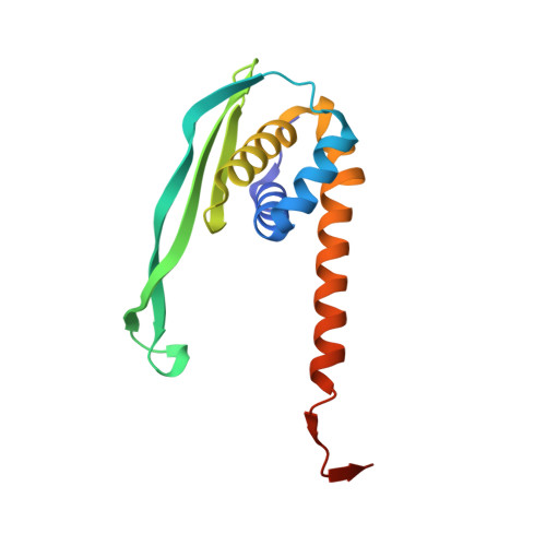
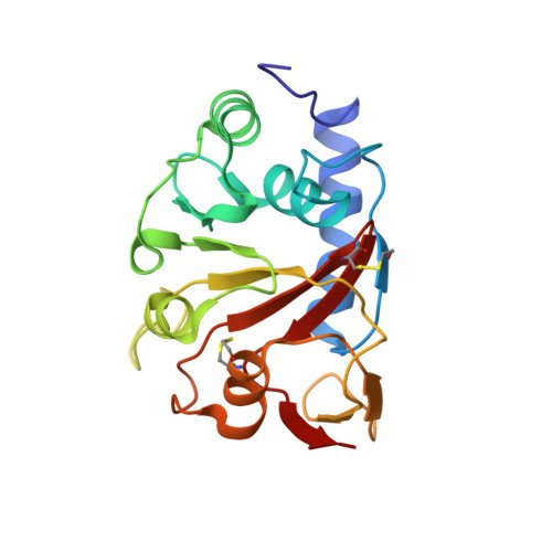
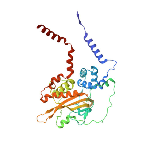
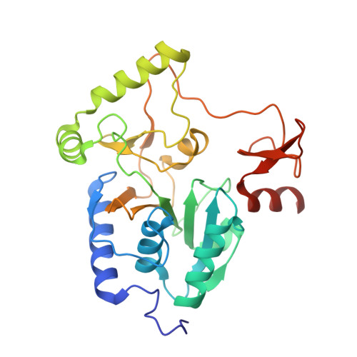
    Phosphorus is required for all life and microorganisms can extract it from their environment through several metabolic pathways. When phosphate is in limited supply, some bacteria are able to use phosphonate compounds, which require specialized enzymatic machinery to break the stable carbon-phosphorus (C-P) bond. Despite its importance, the details of how this machinery catabolizes phosphonates remain unknown. Here we determine the crystal structure of the 240-kilodalton Escherichia coli C-P lyase core complex (PhnG-PhnH-PhnI-PhnJ; PhnGHIJ), and show that it is a two-fold symmetric hetero-octamer comprising an intertwined network of subunits with unexpected self-homologies. It contains two potential active sites that probably couple phosphonate compounds to ATP and subsequently hydrolyse the C-P bond. We map the binding site of PhnK on the complex using electron microscopy, and show that it binds to a conserved insertion domain of PhnJ. Our results provide a structural basis for understanding microbial phosphonate breakdown.


  • Organizational Affiliation

    Department of Molecular Biology and Genetics, Aarhus University, Gustav Wieds Vej 10c, DK-8000 Aarhus C, Denmark.


Macromolecules
Find similar proteins by:  (by identity cutoff)  |  3D Structure
Entity ID: 1
MoleculeChains Sequence LengthOrganismDetailsImage
Alpha-D-ribose 1-methylphosphonate 5-triphosphate synthase subunit PhnG
A, E
150Escherichia coli str. K-12 substr. MG1655Mutation(s): 0 
Gene Names: phnGb4101JW4062
EC: 2.7.8.37
UniProt
Find proteins for P16685 (Escherichia coli (strain K12))
Explore P16685 
Go to UniProtKB:  P16685
Entity Groups  
Sequence Clusters30% Identity50% Identity70% Identity90% Identity95% Identity100% Identity
UniProt GroupP16685
Sequence Annotations
Expand
  • Reference Sequence
Find similar proteins by:  (by identity cutoff)  |  3D Structure
Entity ID: 2
MoleculeChains Sequence LengthOrganismDetailsImage
Alpha-D-ribose 1-methylphosphonate 5-triphosphate synthase subunit PhnH
B, F
194Escherichia coli str. K-12 substr. MG1655Mutation(s): 1 
Gene Names: phnHb4100JW4061
EC: 2.7.8.37
UniProt
Find proteins for P16686 (Escherichia coli (strain K12))
Explore P16686 
Go to UniProtKB:  P16686
Entity Groups  
Sequence Clusters30% Identity50% Identity70% Identity90% Identity95% Identity100% Identity
UniProt GroupP16686
Sequence Annotations
Expand
  • Reference Sequence
Find similar proteins by:  (by identity cutoff)  |  3D Structure
Entity ID: 3
MoleculeChains Sequence LengthOrganismDetailsImage
Alpha-D-ribose 1-methylphosphonate 5-triphosphate synthase subunit PhnI
C, G
354Escherichia coli str. K-12 substr. MG1655Mutation(s): 1 
Gene Names: phnIb4099JW4060
EC: 2.7.8.37
UniProt
Find proteins for P16687 (Escherichia coli (strain K12))
Explore P16687 
Go to UniProtKB:  P16687
Entity Groups  
Sequence Clusters30% Identity50% Identity70% Identity90% Identity95% Identity100% Identity
UniProt GroupP16687
Sequence Annotations
Expand
  • Reference Sequence
Find similar proteins by:  (by identity cutoff)  |  3D Structure
Entity ID: 4
MoleculeChains Sequence LengthOrganismDetailsImage
Alpha-D-ribose 1-methylphosphonate 5-phosphate C-P lyase
D, H
281Escherichia coli str. K-12 substr. MG1655Mutation(s): 0 
Gene Names: phnJb4098JW4059
EC: 4.7.1.1
UniProt
Find proteins for P16688 (Escherichia coli (strain K12))
Explore P16688 
Go to UniProtKB:  P16688
Entity Groups  
Sequence Clusters30% Identity50% Identity70% Identity90% Identity95% Identity100% Identity
UniProt GroupP16688
Sequence Annotations
Expand
  • Reference Sequence
Experimental Data & Validation

Experimental Data

  • Method: X-RAY DIFFRACTION
  • Resolution: 1.70 Å
  • R-Value Free: 0.176 
  • R-Value Work: 0.148 
  • R-Value Observed: 0.150 
  • Space Group: P 21 21 21
Unit Cell:
Length ( Å )Angle ( ˚ )
a = 95.51α = 90
b = 133.71β = 90
c = 176.74γ = 90
Software Package:
Software NamePurpose
PHENIXrefinement
XDSdata reduction
XDSdata scaling
PHASERphasing

Structure Validation

View Full Validation Report


Entry History & Funding Information

Deposition Data


Funding OrganizationLocationGrant Number
Danish National Research FoundationDenmarkCentre for mRNP Biogenesis and Metabolism

Revision History  (Full details and data files)

  • Version 1.0: 2015-08-19
    Type: Initial release
  • Version 1.1: 2015-08-26
    Changes: Database references
  • Version 1.2: 2015-09-09
    Changes: Database references
  • Version 1.3: 2024-01-10
    Changes: Data collection, Database references, Derived calculations, Refinement description