4X8P

Crystal structure of Ash2L SPRY domain in complex with RbBP5


Experimental Data Snapshot

  • Method: X-RAY DIFFRACTION
  • Resolution: 2.20 Å
  • R-Value Free: 0.271 
  • R-Value Work: 0.220 
  • R-Value Observed: 0.222 

wwPDB Validation   3D Report Full Report


This is version 1.1 of the entry. See complete history


Literature

A phosphorylation switch on RbBP5 regulates histone H3 Lys4 methylation.

Zhang, P.Chaturvedi, C.P.Tremblay, V.Cramet, M.Brunzelle, J.S.Skiniotis, G.Brand, M.Shilatifard, A.Couture, J.F.

(2015) Genes Dev 29: 123-128

  • DOI: https://doi.org/10.1101/gad.254870.114
  • Primary Citation of Related Structures:  
    4X8N, 4X8P

  • PubMed Abstract: 

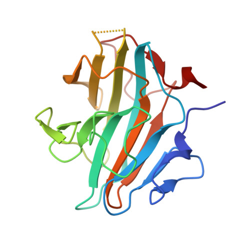
    The methyltransferase activity of the trithorax group (TrxG) protein MLL1 found within its COMPASS (complex associated with SET1)-like complex is allosterically regulated by a four-subunit complex composed of WDR5, RbBP5, Ash2L, and DPY30 (also referred to as WRAD). We report structural evidence showing that in WRAD, a concave surface of the Ash2L SPIa and ryanodine receptor (SPRY) domain binds to a cluster of acidic residues, referred to as the D/E box, in RbBP5. Mutational analysis shows that residues forming the Ash2L/RbBP5 interface are important for heterodimer formation, stimulation of MLL1 catalytic activity, and erythroid cell terminal differentiation. We also demonstrate that a phosphorylation switch on RbBP5 stimulates WRAD complex formation and significantly increases KMT2 (lysine [K] methyltransferase 2) enzyme methylation rates. Overall, our findings provide structural insights into the assembly of the WRAD complex and point to a novel regulatory mechanism controlling the activity of the KMT2/COMPASS family of lysine methyltransferases.


  • Organizational Affiliation

    Department of Biochemistry, Microbiology, and Immunology, Ottawa Institute of Systems Biology, University of Ottawa, Ottawa, Ontario K1H 8M5, Canada;


Macromolecules
Find similar proteins by:  (by identity cutoff)  |  3D Structure
Entity ID: 1
MoleculeChains Sequence LengthOrganismDetailsImage
Set1/Ash2 histone methyltransferase complex subunit ASH2,Set1/Ash2 histone methyltransferase complex subunit ASH2182Homo sapiensMutation(s): 0 
Gene Names: ASH2LASH2L1
UniProt & NIH Common Fund Data Resources
Find proteins for Q9UBL3 (Homo sapiens)
Explore Q9UBL3 
Go to UniProtKB:  Q9UBL3
PHAROS:  Q9UBL3
GTEx:  ENSG00000129691 
Entity Groups  
Sequence Clusters30% Identity50% Identity70% Identity90% Identity95% Identity100% Identity
UniProt GroupQ9UBL3
Sequence Annotations
Expand
  • Reference Sequence

Find similar proteins by:  Sequence   |   3D Structure  

Entity ID: 2
MoleculeChains Sequence LengthOrganismDetailsImage
Retinoblastoma-binding protein 512Homo sapiensMutation(s): 0 
UniProt & NIH Common Fund Data Resources
Find proteins for Q15291 (Homo sapiens)
Explore Q15291 
Go to UniProtKB:  Q15291
PHAROS:  Q15291
GTEx:  ENSG00000117222 
Entity Groups  
Sequence Clusters30% Identity50% Identity70% Identity90% Identity95% Identity100% Identity
UniProt GroupQ15291
Sequence Annotations
Expand
  • Reference Sequence
Small Molecules
Ligands 1 Unique
IDChains Name / Formula / InChI Key2D Diagram3D Interactions
GOL
Query on GOL

Download Ideal Coordinates CCD File 
C [auth B]GLYCEROL
C3 H8 O3
PEDCQBHIVMGVHV-UHFFFAOYSA-N
Experimental Data & Validation

Experimental Data

  • Method: X-RAY DIFFRACTION
  • Resolution: 2.20 Å
  • R-Value Free: 0.271 
  • R-Value Work: 0.220 
  • R-Value Observed: 0.222 
  • Space Group: P 21 21 21
Unit Cell:
Length ( Å )Angle ( ˚ )
a = 51.5α = 90
b = 59.1β = 90
c = 70.8γ = 90
Software Package:
Software NamePurpose
BUSTERrefinement
d*TREKdata reduction
d*TREKdata scaling
PHASERphasing

Structure Validation

View Full Validation Report


Entry History 

Deposition Data

Revision History  (Full details and data files)

  • Version 1.0: 2015-01-28
    Type: Initial release
  • Version 1.1: 2024-02-28
    Changes: Data collection, Database references, Derived calculations, Source and taxonomy