4X7Q

PIM2 kinase in complex with Compound 1s


Experimental Data Snapshot

  • Method: X-RAY DIFFRACTION
  • Resolution: 2.33 Å
  • R-Value Free: 0.258 
  • R-Value Work: 0.196 
  • R-Value Observed: 0.199 

wwPDB Validation   3D Report Full Report


Ligand Structure Quality Assessment 


This is version 1.2 of the entry. See complete history


Literature

Structure-based design of low-nanomolar PIM kinase inhibitors.

Ishchenko, A.Zhang, L.Le Brazidec, J.Y.Fan, J.Chong, J.H.Hingway, A.Raditsis, A.Singh, L.Elenbaas, B.Hong, V.S.Marcotte, D.Silvian, L.Enyedy, I.Chao, J.

(2015) Bioorg Med Chem Lett 25: 474-480

  • DOI: https://doi.org/10.1016/j.bmcl.2014.12.041
  • Primary Citation of Related Structures:  
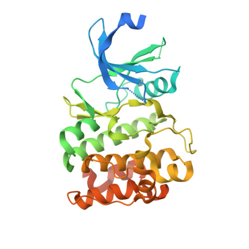
    4X7Q, 4XHK

  • PubMed Abstract: 

    PIM kinases are implicated in variety of cancers by promoting cell survival and proliferation and are targets of interest for therapeutic intervention. We have identified a low-nanomolar pan-PIM inhibitor (PIM1/2/3 potency 5:14:2nM) using structure based modeling. The crystal structure of this compound with PIM1 confirmed the predicted binding mode and protein-ligand interactions except those in the acidic ribose pocket. We show the SAR suggesting the importance of having a hydrogen bond donor in this pocket for inhibiting PIM2; however, this interaction is not important for inhibiting PIM1 or PIM3. In addition, we report the discovery of a new class of PIM inhibitors by using computational de novo design tool implemented in MOE software (Chemical Computing Group). These inhibitors have a different interaction profile.


  • Organizational Affiliation

    Biogen Idec, 14 Cambridge Center, Cambridge, MA 02142, USA. Electronic address: al_ishchenko@yahoo.com.


Macromolecules
Find similar proteins by:  (by identity cutoff)  |  3D Structure
Entity ID: 1
MoleculeChains Sequence LengthOrganismDetailsImage
Serine/threonine-protein kinase pim-2A [auth B],
B [auth A]
312Homo sapiensMutation(s): 0 
Gene Names: PIM2
EC: 2.7.11.1
UniProt & NIH Common Fund Data Resources
Find proteins for Q9P1W9 (Homo sapiens)
Explore Q9P1W9 
Go to UniProtKB:  Q9P1W9
PHAROS:  Q9P1W9
GTEx:  ENSG00000102096 
Entity Groups  
Sequence Clusters30% Identity50% Identity70% Identity90% Identity95% Identity100% Identity
UniProt GroupQ9P1W9
Sequence Annotations
Expand
  • Reference Sequence
Small Molecules
Ligands 2 Unique
IDChains Name / Formula / InChI Key2D Diagram3D Interactions
3YR
Query on 3YR

Download Ideal Coordinates CCD File 
C [auth B],
D [auth A]
2-(2,6-difluorophenyl)-N-{4-[(3S)-pyrrolidin-3-yloxy]pyridin-3-yl}-1,3-thiazole-4-carboxamide
C19 H16 F2 N4 O2 S
DNXZIJKGPFJRAA-NSHDSACASA-N
PO4
Query on PO4

Download Ideal Coordinates CCD File 
E [auth A]PHOSPHATE ION
O4 P
NBIIXXVUZAFLBC-UHFFFAOYSA-K
Binding Affinity Annotations 
IDSourceBinding Affinity
3YR BindingDB:  4X7Q IC50: 14 (nM) from 1 assay(s)
Binding MOAD:  4X7Q IC50: 14 (nM) from 1 assay(s)
Experimental Data & Validation

Experimental Data

  • Method: X-RAY DIFFRACTION
  • Resolution: 2.33 Å
  • R-Value Free: 0.258 
  • R-Value Work: 0.196 
  • R-Value Observed: 0.199 
  • Space Group: H 3
Unit Cell:
Length ( Å )Angle ( ˚ )
a = 142.265α = 90
b = 142.265β = 90
c = 61.75γ = 120
Software Package:
Software NamePurpose
SCALAdata scaling
REFMACrefinement
PDB_EXTRACTdata extraction

Structure Validation

View Full Validation Report


Ligand Structure Quality Assessment 


Entry History 

Deposition Data

Revision History  (Full details and data files)

  • Version 1.0: 2015-02-11
    Type: Initial release
  • Version 1.1: 2017-11-22
    Changes: Database references, Derived calculations, Refinement description, Source and taxonomy
  • Version 1.2: 2023-09-27
    Changes: Data collection, Database references, Refinement description