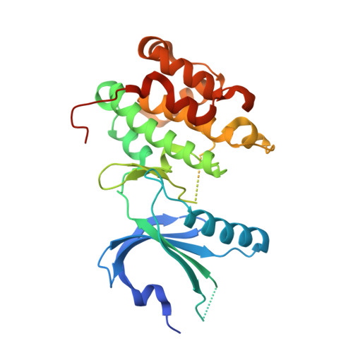
4X7H

Co-crystal Structure of PERK bound to N-{5-[(6,7-dimethoxyquinolin-4-yl)oxy]pyridin-2-yl}-1-methyl-3-oxo-2-phenyl-5-(pyridin-4-yl)-2,3-dihydro-1H-pyrazole-4-carboxamide inhibitor


Experimental Data Snapshot

  • Method: X-RAY DIFFRACTION
  • Resolution: 2.00 Å
  • R-Value Free: 0.214 
  • R-Value Work: 0.201 
  • R-Value Observed: 0.202 

wwPDB Validation   3D Report Full Report


Ligand Structure Quality Assessment 


This is version 1.3 of the entry. See complete history


Literature

Discovery of 1H-Pyrazol-3(2H)-ones as Potent and Selective Inhibitors of Protein Kinase R-like Endoplasmic Reticulum Kinase (PERK).

Smith, A.L.Andrews, K.L.Beckmann, H.Bellon, S.F.Beltran, P.J.Booker, S.Chen, H.Chung, Y.A.D'Angelo, N.D.Dao, J.Dellamaggiore, K.R.Jaeckel, P.Kendall, R.Labitzke, K.Long, A.M.Materna-Reichelt, S.Mitchell, P.Norman, M.H.Powers, D.Rose, M.Shaffer, P.L.Wu, M.M.Lipford, J.R.

(2015) J Med Chem 58: 1426-1441

  • DOI: https://doi.org/10.1021/jm5017494
  • Primary Citation of Related Structures:  
    4X7H, 4X7J, 4X7K, 4X7L, 4X7N, 4X7O

  • PubMed Abstract: 

    The structure-based design and optimization of a novel series of selective PERK inhibitors are described resulting in the identification of 44 as a potent, highly selective, and orally active tool compound suitable for PERK pathway biology exploration both in vitro and in vivo.


  • Organizational Affiliation

    Departments of †Medicinal Chemistry, ‡Molecular Structure and Characterization, §Oncology Research, and ∥Pharmacokinetics and Drug Metabolism, Amgen Inc. , One Amgen Center Drive, Thousand Oaks, California 91320-1799, United States.


Macromolecules
Find similar proteins by:  (by identity cutoff)  |  3D Structure
Entity ID: 1
MoleculeChains Sequence LengthOrganismDetailsImage
Eukaryotic translation initiation factor 2-alpha kinase 3,Eukaryotic translation initiation factor 2-alpha kinase 3317Homo sapiensMutation(s): 1 
Gene Names: EIF2AK3PEKPERK
EC: 2.7.11.1
UniProt & NIH Common Fund Data Resources
Find proteins for Q9NZJ5 (Homo sapiens)
Explore Q9NZJ5 
Go to UniProtKB:  Q9NZJ5
PHAROS:  Q9NZJ5
GTEx:  ENSG00000172071 
Entity Groups  
Sequence Clusters30% Identity50% Identity70% Identity90% Identity95% Identity100% Identity
UniProt GroupQ9NZJ5
Sequence Annotations
Expand
  • Reference Sequence
Small Molecules
Ligands 2 Unique
IDChains Name / Formula / InChI Key2D Diagram3D Interactions
3Z2
Query on 3Z2

Download Ideal Coordinates CCD File 
B [auth A]N-{5-[(6,7-dimethoxyquinolin-4-yl)oxy]pyridin-2-yl}-1-methyl-3-oxo-2-phenyl-5-(pyridin-4-yl)-2,3-dihydro-1H-pyrazole-4-carboxamide
C32 H26 N6 O5
JZUVVTPSJAHDMJ-UHFFFAOYSA-N
SO4
Query on SO4

Download Ideal Coordinates CCD File 
C [auth A],
D [auth A]
SULFATE ION
O4 S
QAOWNCQODCNURD-UHFFFAOYSA-L
Binding Affinity Annotations 
IDSourceBinding Affinity
3Z2 BindingDB:  4X7H IC50: 42 (nM) from 1 assay(s)
Experimental Data & Validation

Experimental Data

  • Method: X-RAY DIFFRACTION
  • Resolution: 2.00 Å
  • R-Value Free: 0.214 
  • R-Value Work: 0.201 
  • R-Value Observed: 0.202 
  • Space Group: P 41 21 2
Unit Cell:
Length ( Å )Angle ( ˚ )
a = 81.428α = 90
b = 81.428β = 90
c = 127.933γ = 90
Software Package:
Software NamePurpose
REFMACrefinement
SCALEPACKdata scaling
PDB_EXTRACTdata extraction
MOLREPphasing

Structure Validation

View Full Validation Report


Ligand Structure Quality Assessment 


Entry History 

Deposition Data

Revision History  (Full details and data files)

  • Version 1.0: 2015-02-04
    Type: Initial release
  • Version 1.1: 2015-02-25
    Changes: Database references
  • Version 1.2: 2017-11-22
    Changes: Database references, Derived calculations, Refinement description, Source and taxonomy, Structure summary
  • Version 1.3: 2023-09-27
    Changes: Data collection, Database references, Refinement description