Structural basis of Dscam1 homodimerization: Insights into context constraint for protein recognition
Li, S.A., Cheng, L., Yu, Y., Chen, Q.(2016) Sci Adv 2: e1501118-e1501118
- PubMed: 27386517
- DOI: https://doi.org/10.1126/sciadv.1501118
- Primary Citation of Related Structures:
4WVR, 4X5L, 4X83, 4X8X, 4X9B, 4X9F, 4X9G, 4X9H, 4X9I, 4XB7, 4XB8, 4XHQ - PubMed Abstract:
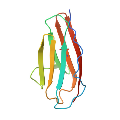
The Drosophila neural receptor Dscam1 (Down syndrome cell adhesion molecule 1) plays an essential role in neuronal wiring and self-avoidance. Dscam1 potentially encodes 19,008 ectodomains through alternative RNA splicing and exhibits exquisite isoform-specific homophilic binding, which makes it an exceptional example for studying protein binding specificity. However, structural information on Dscam1 is limited, which hinders illumination of the mechanism of Dscam1 isoform-specific recognition. Whether different Dscam1 isoforms adopt the same dimerization mode remains a subject of debate. We present 12 Dscam1 crystal structures, provide direct evidence indicating that all isoforms adopt a conserved homodimer geometry in a modular fashion, identify two mechanisms for the Ig2 binding domain to dispel electrostatic repulsion during dimerization, decode Ig2 binding specificity by a central motif at its symmetry center, uncover the role of glycosylation in Dscam1 homodimerization, and find electrostatic potential complementarity to help define the binding region and the antiparallel binding mode. We then propose a concept that the context of a protein may set restrictions to regulate its binding specificity, which provides a better understanding of protein recognition.
Organizational Affiliation:
State Key Laboratory of Biotherapy and Cancer Center, West China Hospital, Sichuan University, and Collaborative Innovation Center of Biotherapy, Chengdu 610041, P. R. China.